October 11th, 2022
Written by: Joe Stucynski
Imagine you’re a firefighter at work. Occasionally the alarm will sound and it’s all hands on deck as you race to the fire to put it out. But what do you do when there’s no fire? Well you can do a lot of things. There’s equipment to clean and maintain. You need to make sure the fire truck’s ready to go at a moment’s notice. Maybe the firehouse needs to be painted again. Or maybe you put together some community building events to help the neighborhood. Whatever it is, just because there’s no emergency doesn’t mean there aren’t things to do.
When it comes to the brain, cells called microglia are like the firefighters: always active and ready to address problems. Because immune cells in the body can’t easily cross into the brain, microglia fill that role and patrol the whole brain, watching out for emergencies and responding to them when they find one. Neuroscientists estimate that up to 10% of all cells in the brain are microglia!1 Most past research has studied how microglia respond in emergencies, rushing to sites of neural injury, hunting down and killing foreign bacteria, targeting inflammation, and clearing dead cells and debris. Much less research has addressed what they do in the healthy brain and how they might influence brain activity when they’re not responding to emergencies.
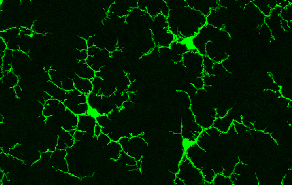
When microglia are ‘resting’ they are much smaller than when they’re responding to emergencies, but they actively reach out long arms to their surroundings to sense molecular signals coming from anything nearby that is going wrong. Conversely, when they sense a problem to address, they switch modes to become much larger, their probing arms shrink, and they travel to the location of the emergency. For a more detailed explanation, see the following video.
While neuroscientists initially thought that microglia are mostly passive when extending their arms to detect an emergency, they may actually play a more active role in regulating the brain than previously thought. When microglia actively reach out, they can make contact with nearby connections between neurons. By doing this they can affect the connection strength or even remove weak links.1 Neuroscientists have shown that when microglia contact neurons this way they can affect social behavior, spatial learning, and memory.1 In a healthy brain, microglia also help you forget irrelevant information which is good, but appear to malfunction in Alzheimer’s disease contributing to loss of important memories.
It has also recently been shown that microglia switch between their two activity modes during sleep versus wakefulness. During sleep, microglia are much more likely to investigate their local environment than they are during wake. They look for emergencies and touch neurons to check on connections and adjust them. There are several mechanisms that might explain this shift during sleep. For instance, several recent studies showed that microglia have receptors for a signaling molecule called norepinephrine. When norepinephrine levels are high during wake, it causes microglia to retract their branches, whereas when norepinephrine levels are low during sleep, microglia can survey their environment more readily.2,3 Another study showed that sleep/wake differences in microglia activity depend on the circadian rhythm, a robust 24 hour molecular clock in the brain that helps tell the brain when to be awake or asleep to better match the time of day. This study showed that knocking out some of the core genes that make the circadian clock function correctly prevented the daily differences in microglia activity during sleep versus wake,4 indicating that the molecular clock directly controls microglia rhythms.
These studies showed that sleep and wake can affect the activity of microglia, but how about the opposite? Can the activity of microglia affect how much sleep you get? The answer appears to be yes. By reaching out and contacting connections between neurons during sleep, microglia are able to increase the level of neural synchronization in a population of neurons.5 Increased neural synchronization is a hallmark of brain activity during sleep, which means microglia may therefore promote deeper sleep.
Sleep is synchronized over the whole brain by a network of neuronal populations that act as switches: when the network turns on, the brain is awake, but when the network turns off the brain is asleep. By specifically influencing the neurons in this sleep/wake network, microglia could have a big impact on your quality of sleep. For instance, a recent study found that several types of waste products produced by neurons during wake affect how well neurons in the sleep network can communicate. The neuroscientists showed that microglia are critical to removing those waste products and adjusting communications between the neurons controlling sleep so that normal amounts of sleep could occur.6 These findings may be relevant to patients with Alzheimer’s disease who suffer from severe sleep disruptions, build-up of neuronal waste products, and altered microglia functioning.7,8
All in all, microglia are vital to normal brain function, even in the absence of neural injury or external threats. As their mysteries begin to unravel, and we learn just how much they shape brain activity, we just might have to redefine what it means for microglia to be at ‘rest’.
References
- Deurveilher, S., Golovin, T., Hall, S., Semba, K. Microglia Dynamics in sleep/wake state and in response to sleep loss. Neurochemistry International, 2021.
- Stowell, R.D., Sipe, G.O., Dawes, R.P., Batchelor, N.H., Lordy, K.A., Whitelaw, B.S., Stoessel, MB., Bidlack, J.M., Brown, E., Sur, M., Majewska, A.K. Noradrenergic signaling in the wakeful state inhibits microglial surveillance and synaptic plasticity in the mouse visual cortex. Nature Neuroscience, 2019.
- Liu, Y.U., Uing, Y., Li, Y., Eo, U.B., Chen, T., Zheng, J., Umpierre, A.D., Zhu, J., Bosco, D.B., Dong, H., Wu, LJ. Neuronal network activity controls microglial process surveillance in awake mice via norepinephrine signaling. Nature Neuroscience, 2019.
- Hayashi, Y., Koyanagi, S., Kusunose, N., Takayama, F., Okada, R., Wu, Z., Ohdo, S., Nakanishi, H. Diurnal spatial rearrangement of microglial process through the rhythmic expression of P2Y12 receptors. Neurological Disorders, 2013.
- Akiyoshi, R., Wake, H., Kato, D., Horiuchi, H., Ono, R., Ikegami, A., Haruwaka, K., Omori, T., Tachibana, Y., Moorhouse, A.J., Nabekura, J. Microglia enhance synapse activity to promote local network synchronization. eNeuro, 2018.
- Liu, H., Wang, X., Chen, L., Chen, L., Tsirka, S.E., Ge, S., Xiong, Q. Microglia modulate stable wakefulness via the thalamic reticular nucleus in mice. Nature Communications, 2021.
- Filippov, V., Song, M.A., Zhang, K., Vinters, H.V., Tung, S., Kirsch, W.M., Yang, J., Duerksen-Hughes, P.J. Increased ceramide in brains with Alzheimer’s and other neurodegenerative diseases. Journal of Alzheimer’s Disease, 2012.
- Vitiello, M.V., Borson, S., Sleep disturbance in patients with Alzheimer’s disesase. Mol Diag Ther, 2001.
Cover image by Wai T. Wong, National Eye Institute, NIH via Wikimedia Commons.

.jpg)