April 9th, 2024
Written by: Julia Riley
When we talk about neuroscience research, cells called neurons are usually the main character. Neurons have the ability to transmit electrical currents, and this unique form of communication ultimately gives rise to our ability to function. As such, many scientists are interested in clarifying how neurons communicate and what influences their communication. Despite their main character-type status in neuroscience, neurons are far from the only type of cells in the brain- in fact, they aren’t even the most numerous!1
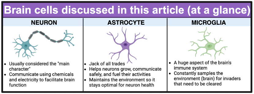
Around the same time neurons were discovered, another class of cells called glia were also described.2 Their namesake is the Greek word for glue; at the time, they were thought to be simple supporting character-type cells with the sole purpose of holding neurons in place.3 We’ve since learned that there are several types of glia, and each is uniquely qualified to play their own specific and important role in brain health and disease.1,3 We also now understand that neurons and glia are constantly communicating with each other; if one population of cells goes haywire, the others could very realistically follow.4 However, because investigation into these types of brain cells was neglected for so long, we still have a lot to learn about how each of these cell types function normally, let alone how they might act differently in the context of certain diseases.
One group of diseases where the contributions of glia are beginning to be investigated are neurodegenerative diseases. Neurodegenerative diseases are a group of diseases caused by the irreversible death of neurons. Some examples of neurodegenerative diseases include Parkinson’s, Alzheimer’s, and amyotrophic lateral sclerosis (also known as ALS or Lou Gehrig’s disease).5 Since neurons continuously die and cannot be replenished in neurodegenerative diseases, the symptoms that patients experience consistently worsen with time.5 While neurons have been the main focus of most neurodegenerative disease-oriented research, studies are beginning to suggest that glial cells might play an active role in the progression of these diseases.1,6,7 It remains unclear whether that role is protective or harmful toward neuron health, or if it differs on a case-by-case basis.
Here, we’re going to dive into two of the major glial cell types in the brain (Figure 1), their day-to-day jobs, and some current thoughts on how they might contribute to worsening or improving neurodegenerative diseases.
Astrocytes
Astrocytes are glial cells named for their star-like or stellate shape8. Appropriately, they are absolute stars when it comes to influencing brain health! Astrocytes play critical roles in making sure neurons exist in an environment that allows them to function optimally. This includes supplying neurons with molecules they can turn into energy,8 maintaining the environment in the brain so neurons can communicate properly,9 and helping neurons form connections with one another throughout your life (Figure 2).10
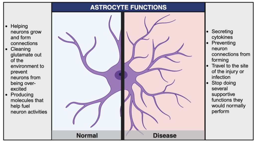
One of the major jobs of astrocytes is to maintain the environment in the brain so that neurons can function. Because of this, astrocytes are phenomenal at sensing and responding to cues in their environment. For example, if an astrocyte senses the presence of something dangerous that shouldn’t be in the brain, it might secrete chemicals called cytokines.6,11 Cytokines are important molecular messengers for the process of inflammation. While you might think of inflammation as swelling that happens after an injury, it is actually a catch-all phrase for a variety of methods that the body uses to indicate that something is wrong so that it can prepare to defend itself. This is most frequently triggered by foreign invaders entering the body, which can be anything from bacteria or viruses to a malfunctional or damaged molecule produced by an unhealthy cell in the environment. This means that in addition to the type of large-scale inflammation you can see with the naked eye (like swelling), inflammation also happens on a cellular level, and serves as a signal to other cells that there is a problem in a specific location that needs to be addressed. This is where cytokines come into play. Secretion of cytokines serves a multitude of functions, including calling certain cells to the site of the issue such that it can be delt with in a timely manner. They can also act as an environmental cue to trigger a change in astrocyte behavior. When an astrocyte has started acting differently than it normally would in order to respond to something in its environment, it is called a reactive astrocyte.11 Possibly one of the most interesting aspects about the ability of astrocytes to become reactive is that they seem to truly react in specific ways to specific stimuli.11 Just like you might react to a fire by calling the fire department or react to a bank robbery by calling the police, reactive astrocytes come in many forms that have various consequences for the brain’s health, most of which have yet to be characterized.
Although astrocyte reactivity can be helpful as an initial response when the brain is threatened by environmental factors, long-term exposure to cytokines and other inflammatory molecules can be harmful for a number of reasons. These include causing an inflammatory reaction in other cell types, which could result in their eventual death.12 This would be akin to a tiny fire starting in your kitchen, and you continuing to spray a fire extinguisher until long after the fire was gone- you would probably do as much or more damage to your kitchen with your reaction as was done by the original fire. In several neurodegenerative diseases, we suspect astrocytes may be exacerbating the problem by carrying out an inappropriately prolonged inflammatory response, resulting in damage to nearby cells.
It is also possible that astrocytes cease to perform their helpful functions when in certain reactive states. If this is the case, they could be damaging neurons simply by removing a support system that neurons need to thrive. In some neurodegenerative diseases, it has been shown that astrocytes cease to support connections between neurons necessary for them to communicate. They can also stop cleaning up a molecule called glutamate as efficiently as normal.13 Some neurons secrete glutamate as a method of communication with other neurons (you can read more about glutamate here). However, much like you might get overstimulated if someone is constantly talking at you for far too long, neurons become overexcited when glutamate is not removed from the environment efficiently by astrocytes.14 This can result in neurons dying. These are just a few examples of astrocyte functions that can be altered in disease. Astrocytes in an unhealthy brain might also promote damage by conducting excessive inflammatory communication with microglia, another glial cell type.
Microglia
The immune system that protects the majority of your body from harmful molecules (also called pathogens) cannot get into the brain’s environment under normal circumstances.15,16 This is because the brain is such a unique and delicate organ that it could sustain damage if it was constantly being invaded by cells that weren’t intended to function there. As such, the human body developed a specific glial cell type to serve as the brain’s own special immune system. As the resident immune cells in the brain, microglia are the cell type that most commonly respond to pathogens that try to invade the brain.16
Microglia have the ability to ingest and break down things in the brain that might be harmful, such as bacteria, dead cells, or abnormal clumps of proteins.17 In most neurodegenerative diseases, we see an abnormal buildup of clumps of protein called protein aggregates.18 The exact type of protein that aggregates are made of depends on the disease- for example, in Parkinson’s aggregates are chiefly composed of a protein named alpha-synuclein; in Alzheimer’s, a protein named amyloid-beta steals the show. Microglia can consume and digest protein aggregates in the brain19. This presumably helps to fight disease because it decreases the exposure to protein aggregates inflicted upon neurons.
Like astrocytes, microglia can also become reactive, and are thought to have an especially potent ability to promote inflammation when reactive in response to certain environmental cues.19 One context in which this occurs is during ageing. As we age, inflammation tends to increase throughout the body.20 This is important in the context of neurodegenerative diseases in that most of these diseases are ageing-related; increasing age is the greatest predictor of whether someone will develop Parkinson’s or Alzheimer’s, and you rarely see these diseases in people under 40. We do not yet know why these diseases are more common in older people, but scientists think it might have something to do with the increasing inflammation in the body. making it more vulnerable to developing these diseases with age. One recent study showed that with age, microglia activate at least one specific inflammatory pathway.21 This behavior in microglia is sufficient (capable in and of itself) to cause the inflammation-related changes we see in mice as they age.21 The authors of this study suggest that this increase in inflammation propagated by microglia might be one factor that sets the stage for the development of neurodegenerative disease.
Reactive microglia also have certain tasks they can perform much more frequently and efficiently than they would normally. One task microglia perform is phagocytosis, or the ability to ingest free-floating pathogens or other damaging agents and digest them to exterminate them from the brain.17,19 In certain reactive states, microglia produce more of the molecular tools that allow them to digest things, thus presumably allowing for more efficient clearance of pathogens.19 Some types of reactive microglia can also produce cytokines, and secretion of these cytokines can set off an inflammatory response at the site of brain injury, much like astrocytes can. While it remains unclear where the line between the helpful and hurtful behaviors of reactive microglia lies, we have made great strides in understanding how these cells function in different contexts. Currently, scientists are continuing to investigate basic questions about microglial function, while also exploring ways they could potentially be utilized to help combat disease.
Conclusion
The onset and progression of neurodegenerative diseases is extremely complex, and part of the reason that we don’t understand how to treat them yet is that we don’t understand what causes them in the first place. Besides the decline in heath and eventual death observed in neurons, we have abundant evidence to believe that other cell types like astrocytes and microglia are also contributing to these illnesses. There are other glial cell types whose potential contributions to the development of neurodegenerative diseases are also important and under investigation. A better understanding of how non-neuronal cells contribute to neurodegenerative diseases could ultimately help us disentangle a more efficient way to treat them.
———
If you’d like to read more about these cell types in related PennNeuroKnow posts, you can read about astrocytes here and microglia here.
References
1. Sofroniew, M. V. & Vinters, H. V. Astrocytes: biology and pathology. Acta Neuropathol. (Berl.) 119, 7–35 (2010).
2. Ndubaku, U. & de Bellard, M. E. Glial cells: Old cells with new twists. Acta Histochem. 110, 182–195 (2008).
3. Allen, N. J. & Lyons, D. A. Glia as Architects of Central Nervous System Formation and Function. Science 362, 181–185 (2018).
4. Patel, D. C., Tewari, B. P., Chaunsali, L. & Sontheimer, H. Neuron–glia interactions in the pathophysiology of epilepsy. Nat. Rev. Neurosci. 20, 282–297 (2019).
5. Lamptey, R. N. L. et al. A Review of the Common Neurodegenerative Disorders: Current Therapeutic Approaches and the Potential Role of Nanotherapeutics. Int. J. Mol. Sci. 23, 1851 (2022).
6. Gleichman, A. J. & Carmichael, S. T. Glia in neurodegeneration: Drivers of disease or along for the ride? Neurobiol. Dis. 142, 104957 (2020).
7. Stevenson, R., Samokhina, E., Rossetti, I., Morley, J. W. & Buskila, Y. Neuromodulation of Glial Function During Neurodegeneration. Front. Cell. Neurosci. 14, (2020).
8. Parpura, V. & Verkhratsky, A. Astrocytes revisited: concise historic outlook on glutamate homeostasis and signaling. Croat. Med. J. 53, 518–528 (2012).
9. Bylicky, M. A., Mueller, G. P. & Day, R. M. Mechanisms of Endogenous Neuroprotective Effects of Astrocytes in Brain Injury. Oxid. Med. Cell. Longev. 2018, 6501031 (2018).
10. Molofsky, A. V. et al. Astrocytes and disease: a neurodevelopmental perspective. Genes Dev. 26, 891–907 (2012).
11. Escartin, C. et al. Reactive astrocyte nomenclature, definitions, and future directions. Nat. Neurosci. 24, 312–325 (2021).
12. Zamanian, J. L. et al. Genomic Analysis of Reactive Astrogliosis. J. Neurosci. 32, 6391–6410 (2012).
13. Phatnani, H. & Maniatis, T. Astrocytes in Neurodegenerative Disease. Cold Spring Harb. Perspect. Biol. 7, a020628 (2015).
14. Zhang, L.-N. et al. Astrocytes enhance the tolerance of rat cortical neurons to glutamate excitotoxicity. Mol. Med. Rep. 19, 1521–1528 (2019).
15. Winkler, B. et al. Brain inflammation triggers macrophage invasion across the blood-brain barrier in Drosophila during pupal stages. Sci. Adv. 7, eabh0050.
16. Corraliza, I. Recruiting specialized macrophages across the borders to restore brain functions. Front. Cell. Neurosci. 8, (2014).
17. Lannes, N., Eppler, E., Etemad, S., Yotovski, P. & Filgueira, L. Microglia at center stage: a comprehensive review about the versatile and unique residential macrophages of the central nervous system. Oncotarget 8, 114393–114413 (2017).
18. Ross, C. A. & Poirier, M. A. Protein aggregation and neurodegenerative disease. Nat. Med. 10, S10–S17 (2004).
19. Gao, C., Jiang, J., Tan, Y. & Chen, S. Microglia in neurodegenerative diseases: mechanism and potential therapeutic targets. Signal Transduct. Target. Ther. 8, 1–37 (2023).
20. Li, X. et al. Inflammation and aging: signaling pathways and intervention therapies. Signal Transduct. Target. Ther. 8, 1–29 (2023).
21. Gulen, M. F. et al. cGAS–STING drives ageing-related inflammation and neurodegeneration. Nature 620, 374–380 (2023).
Figures 1 and 2: Created by Julia Riley in Biorender. Publication and Licensing rights agreement number TW26NCMR87.
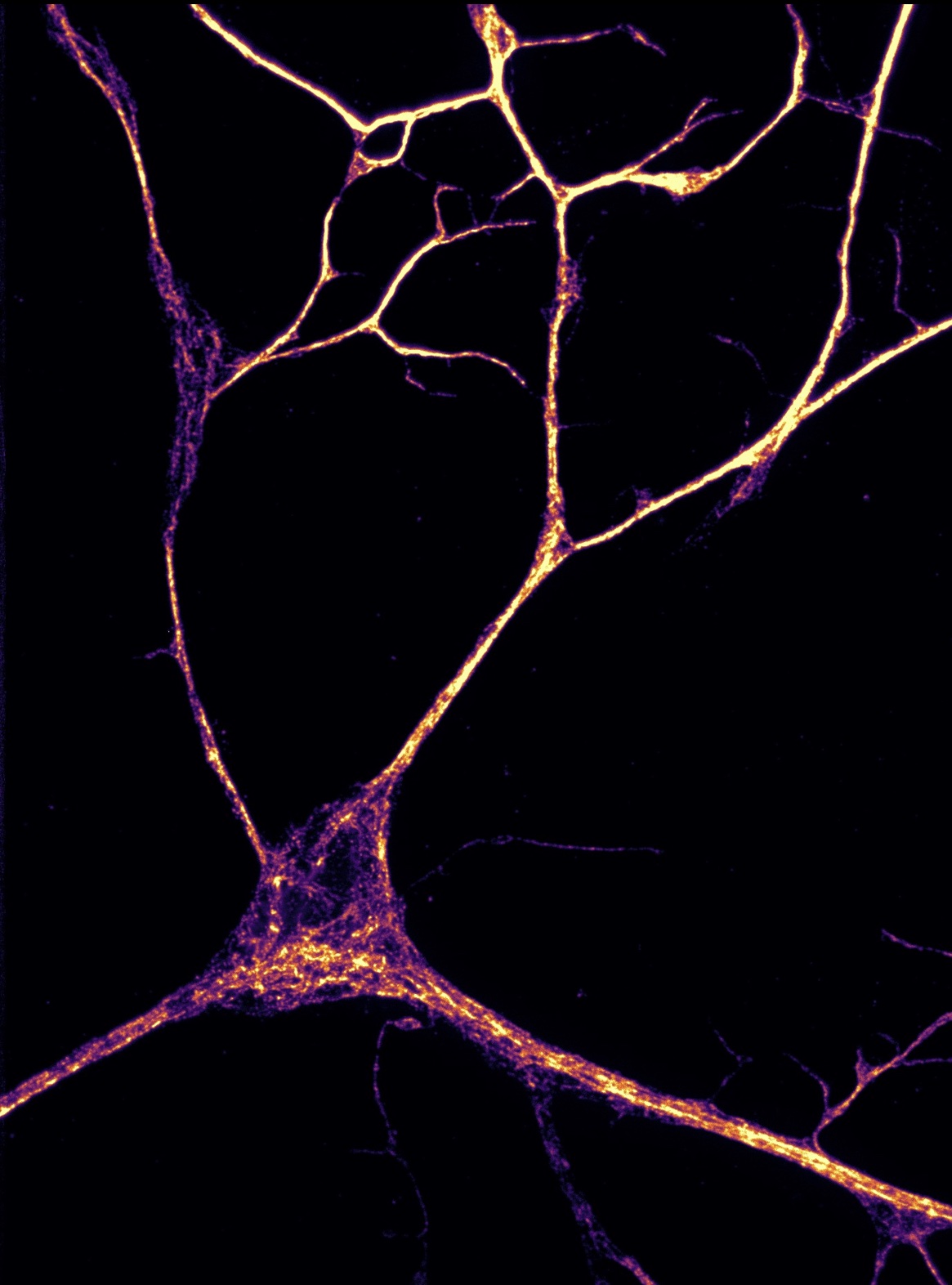
Cover image: Acquired by author Julia Riley for her PhD research.

Leave a comment