October 4th, 2022
Written by: Morgan Kindel
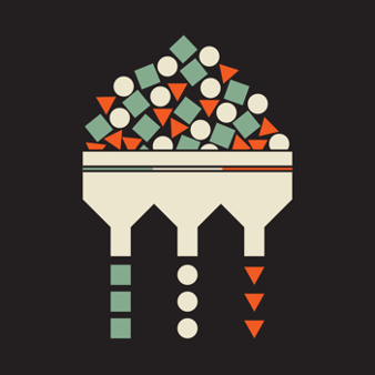
If you’ve ever tried to organize your closet, you know the struggle of choosing how to categorize your clothing items. You could organize by visual cues, like color, style, or pattern, but this isn’t always the most practical, as it overlooks the purpose of each item. A perhaps more practical way to categorize would be by function: item type, warmth, length, etc. The issue of categorization is the same one that neuroscientists commonly face when trying to categorize neurons in the brain. Why is trying to categorize hundreds of billions of neurons important for understanding the brain? To understand why, let’s first review a brief history of neuron categorization.
What we now know as modern-day neuroscience can be traced all the way back to late 19th century Spain, where Santiago Ramón y Cajal, part scientist and part artist, would lock himself in a dark room, crouching over a microscope for hours at a time. Here, he would painstakingly sketch individual neurons, the building blocks of the brain, in intricate detail. These drawings revealed a critical neuroscientific insight: while neurons all possess certain characteristics that enable them to communicate with other cells, they also possess unique shapes and sizes, or, in more scientific terms, morphologies1. Read more about Ramón y Cajal, his drawings, and neuron morphology here!
While morphology may have been the first way to classify neuronal subtypes, it is not the only way. In fact, advancements in molecular biology have inspired a push in neuroscience to define neuronal subpopulations using a different classification method: gene expression. As it turns out, every neuron in the brain has a genetic signature – that is, it expresses a distinct combination of genes, which serve as instruction manuals for the cell that determines its unique characteristics. Neurons that have similar genetic signatures can be grouped together into a group that is sometimes referred to as one type of neuronal population. At the level of an individual neuron, this signature dictates its fundamental properties – not only morphology, but also what other molecules it responds to, and the types of signals that it sends out. When all these neurons come together in a neuronal population, different populations of neurons can produce different widespread brain functions and even different behaviors.
You may have heard that certain brain regions are responsible for different functions. And indeed, brain region is another way to categorize neuronal populations. For example, the amygdala is involved in fear, and the hypothalamus drives hunger. This is not incorrect, but it’s also not the whole picture. Returning to our closet organization analogy, we can organize by item type (shirts, pants, jackets), but even within each of those categories, there will still be differences. We can go further to categorize each clothing type into subcategories (i.e. categorizing shirts into short and long sleeve shirts). We can do something similar in the brain. Within a single brain region, there are many different subpopulations, expressing different genes that have completely different functions2!
One example of this is in a subregion of the hypothalamus called the arcuate nucleus, or Arc, for short. In the Arc, one subpopulation of neurons is activated when we are hungry and drives us to eat3. On the other hand, a different subpopulation within the same region is activated when we are full and drives us to stop eating4. So, while it is true that the Arc drives hunger, it is also true that the Arc suppresses hunger. This contradictory statement can be reconciled by the understanding that there are two different populations of neurons within the Arc, controlling an entirely opposite set of functions.
If you still aren’t totally convinced that studying these distinct neuronal populations is worthwhile, consider this: many psychiatric medications are riddled with side effects that significantly impair quality of life. Some of these side effects are so severe, in fact, that individuals opt out of taking the medication entirely, despite being effective to treat the disorder. Many of these side effects are due to non-specificity of the medication. A medication that blocks a type of dopamine receptor, for example, will block all these dopamine receptors in the brain, not just on the neurons where they are dysfunctional. Side effects arise from blocking the dopamine receptors that are the same type as the dysfunctional ones, but contribute to other, unrelated brain functions. If a specific neuronal subpopulation(s) involved in the disorder were to be identified, then medications could be developed to act only on the dopamine receptors on those dysfunctional neurons, leaving the functioning neurons unbothered, and drastically reducing the number of side effects.
Due to the invasive nature of available technologies used to study genetically defined neuronal populations, so far this work has primarily been done in rodents. As a result, whether the same genetically identified neuronal populations function similarly in human brains isn’t totally clear. To address this, the National Institutes of Health recently announced several initiatives to help fund this research, including one called the BRAIN Initiative Cell Atlas Network which aims to create a map of the different cell types in the human brain. So, while there is still work to be done, considering this path of discovery started out with some sketches, we are well on our way.
References
1. Jiang, X. et al. Principles of connectivity among morphologically defined cell types in adult neocortex. Science 350, aac9462 (2015).
2. Zeng, H. & Sanes, J. R. Neuronal cell-type classification: challenges, opportunities and the path forward. Nat. Rev. Neurosci. 18, 530–546 (2017).
3. van den Top, M., Lee, K., Whyment, A. D., Blanks, A. M. & Spanswick, D. Orexigen-sensitive NPY/AgRP pacemaker neurons in the hypothalamic arcuate nucleus. Nat. Neurosci. 7, 493–494 (2004).
4. Cowley, M. A. et al. Leptin activates anorexigenic POMC neurons through a neural network in the arcuate nucleus. Nature 411, 480–484 (2001).
Cover Image by Igor Kisselev on Adobe Stock

Leave a comment