July 5th, 2022
Written by: Margaret Gardner
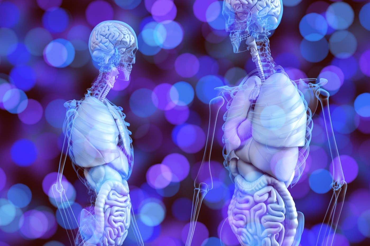
If you’ve heard of glutamate, it’s probably thanks either to its role as the brain’s main excitatory signaling molecule or for putting the “G” in MSG. In both health and disease, glutamate is responsible for a wide variety of functions throughout our bodies and across our lifespan. This week, we’ll take a dive into a few of glutamate’s most prominent roles in the brain.
Neurotransmission
The most famous of glutamate’s uses is as a neurotransmitter, or signaling molecule sent from one neuron to the next at connection points called synapses. It can be found at 1/3 of the brain’s synapses1, and with over 20 types of glutamate receptors in mammals2, it can have a wide range of effects on the cells that receive it. Glutamate receptors can belong to one of two groups: ionotropic and metabotropic1,3.
Ionotropic glutamate receptors have a channel built in, so once glutamate binds, they open up to let certain positive molecules to enter the cell3, much like using a key to open a door. Allowing in positively charged ions is essential to glutamate’s most celebrated job, excitatory signaling. Your brain keeps different amounts of positive and negative molecules inside and outside neurons so that most of the time, neurons are a little bit more negative than the things around them. This works just like a battery with the plus and minus end: in this case the minus end is the neuron, and the plus end is all the other cells and liquid in the brain. So, when glutamate binds, letting in positively charged ions like sodium and calcium, it’s like connecting the plus and minus ends of a battery with wire; the power flows through the path of the wire (or ion channel), evening out the charges across the battery (or raising the neuron’s electrical charge). Raising the neuron’s voltage activates – or excites – it to send a sign to other neurons 3. Glutamate’s metabotropic receptors, meanwhile, trigger slow pathways that can regulate this excitatory signaling, as well as change protein production or the amount of neurotransmitter released2,4.
Glutamate’s excitatory action is important not only for sending immediate information between neurons but also for adjusting the strength of these signaling connections. Since it can strengthen important, commonly used pathways and weaken others as needed, glutamate signaling is the foundation of how the brain learns and remembers5 (two things yours is hopefully doing right now). Disorders such as Alzheimer’s disease seem to attack these glutamate receptors specifically, which may be what causes patients’ impairments in memory and learning4.
While this excitatory activity is necessary for healthy brain function, it is also possible for cells to become overexcited. If glutamate receptors are over-activated and allow too many positive ions into the neuron, the cell will realize something is terribly wrong and activate enzymes causing self-destruction6. This process, known as excitotoxicity, can happen quickly when the brain is hurt, such as by a traumatic brain injury or stroke1,4. Interestingly, it is also thought to play a role in long-term, degenerative brain diseases like amyotrophic lateral sclerosis (ALS), Alzheimer’s, Huntington’s and Parkinson’s diseases1,4,7. Similarly, high glutamate levels can create a vicious cycle where excitation causes seizures that further increase glutamate and lead to excitotoxic damage, perpetuating more seizures9. Though glutamate’s role in epilepsy is complex8, treatments like ketamine, an ionotropic glutamate receptor blocker, and the ketogenic (or “keto”) diet, which triggers the body to make its own ionotropic glutamate receptor blockers, can control seizures by normalizing overactive glutamate signals8.
Regulating Other Neurotransmitters
As a large contributor to the brain’s overall chemical signaling, glutamate affects many other neurotransmitter systems. Glutamate is in fact directly converted to make GABA, the brain’s principal inhibitory neurotransmitter and glutamate’s opposite10; while glutamate’s ionotropic receptors raise neurons’ voltage and make them fire a signal, GABA’s receptors lower neuron’s voltages and keep them quiet. Basically, if glutamate is a car’s gas pedal, GABA is the brakes, and any car worth driving is going to need both. This car is unique, though, in that new brakes can only be made from old engine parts, so changes in glutamate will also affect how much GABA the brain can make.
Many studies have also explored glutamate’s influence on dopamine, particularly in relation to schizophrenia11. Researchers have long thought that problems with dopamine signals could lead to schizophrenia. However, after noticing how well ketamine can recreate the symptoms of schizophrenia in healthy people, scientists have found increasing evidence that reduced ionotropic glutamate receptor activation – which leads to increased dopamine and other abnormalities – may cause the disease, rather than changes in dopamine only11.
Glial Signaling and Cycling
In addition to signaling to neurons, glutamate also interacts with the brain’s white matter, or glial cells, in a number of different ways. Glial cells are a family of fatty white cells in the brain that do everything from insulate neurons’ electrical wiring to coordinate immune function. Like neurons, different varieties of glia also have different types of glutamate receptors, as well as transporters for pulling glutamate into the cell4.
Glia use these glutamate transporters in their role as recyclers; because too much glutamate can cause excitotoxicity, glia have the crucial job of snatching up the glutamate that neurons release, inactivating it, then safely returning it for re-activation and re-use10. This cycle is so critical that up to 80% of the brain’s total glucose is used to power it12. However, changes in the brain’s chemical balance, stroke, or tumors can cause glia to dump their glutamate back onto neurons, triggering excitotoxicity1,2. In multiple sclerosis (MS) and in white-matter tumors, glia struggle to take in glutamate1,13, and disruption of this cycle is implicated in seizures, depressive symptoms, and ALS2,8.
As you can see, glutamate’s to-do list is already quite long, and it’s only getting started. Stay tuned for Part 2, coming soon to a neuroscience blog near you!
References
1. Tapiero H, Mathé G, Couvreur P, Tew KD. II. Glutamine and glutamate. Biomedicine & pharmacotherapy = Biomedecine & pharmacotherapie. 2002;56(9):446-457. doi:10.1016/S0753-3322(02)00285-8
2. Pankevich DE, Davis M, Altevogt BM. GLUTAMATE-RELATED BIOMARKERS IN DRUG DEVELOPMENT FOR DISORDERS OF THE NERVOUS SYSTEM WORKSHOP SUMMARY Forum on Neuroscience and Nervous System Disorders Board on Health Sciences Policy. The National Academies Press; 2011. Accessed June 23, 2022. http://www.nap.edu.
3. Brosnan JT, Brosnan ME. Glutamate: a truly functional amino acid. Amino Acids. 2013;45(3):413-418. doi:10.1007/S00726-012-1280-4
4. Crupi R, Impellizzeri D, Cuzzocrea S. Role of metabotropic glutamate receptors in neurological disorders. Frontiers in Molecular Neuroscience. 2019;12:20. doi:10.3389/FNMOL.2019.00020/BIBTEX
5. Collingridge GL, Bliss TVP. NMDA receptors – their role in long-term potentiation. Trends in Neurosciences. 1987;10(7):288-293. doi:10.1016/0166-2236(87)90175-5
6. Meldrum BS. Glutamate as a Neurotransmitter in the Brain: Review of Physiology and Pathology. The Journal of Nutrition. 2000;130(4):1007S-1015S. doi:10.1093/JN/130.4.1007S
7. Zhang X, Wang D, Zhang B, Zhu J, Zhou Z, Cui L. Regulation of microglia by glutamate and its signal pathway in neurodegenerative diseases. Drug Discov Today. 2020;25(6):1074-1085. doi:10.1016/J.DRUDIS.2020.04.001
8. Eid T, Gruenbaum SE, Dhaher R, Lee TSW, Zhou Y, Danbolt NC. The glutamate-glutamine cycle in epilepsy. In: The Glutamate/GABA-Glutamine Cycle. Vol 13. Springer Science and Business Media, LLC; 2016:351-400. doi:10.1007/978-3-319-45096-4_14/FIGURES/5
9. Barker-Haliski M, Steve White H. Glutamatergic Mechanisms Associated with Seizures and Epilepsy. Cold Spring Harbor Perspectives in Medicine. 2015;5(8):1-15. doi:10.1101/CSHPERSPECT.A022863
10. Petroff OAC. GABA and glutamate in the human brain. Neuroscientist. 2002;8(6):562-573. doi:10.1177/1073858402238515
11. Poels E, Kegeles LS, Kantrowitz JT, et al. Imaging glutamate in schizophrenia: review of findings and implications for drug discovery. Molecular Psychiatry. 2014;19:20-29. doi:10.1038/mp.2013.136
12. Rothman DL, Behar KL, Hyder F, Shulman RG. In vivo NMR studies of the glutamate neurotransmitter flux and neuroenergetics: implications for brain function. Annu Rev Physiol. 2003;65:401-427. doi:10.1146/ANNUREV.PHYSIOL.65.092101.142131
13. Spitzer S, Volbracht K, Lundgaard I, Káradóttir RT. Glutamate signalling: A multifaceted modulator of oligodendrocyte lineage cells in health and disease. Neuropharmacology. 2016;110(Pt B):574-585. doi:10.1016/J.NEUROPHARM.2016.06.014
