December 4, 2018
Written by: Nitsan Goldstein
Chances are you know someone that suffers or has suffered from a psychiatric disorder such as depression or a neurodegenerative disease like Alzheimer’s or Parkinson’s diseases. If that is the case, you know there are medications and other therapies out there, but there is no cure for these and most other neurological diseases. Why is this? One major reason is that we are highly limited in our ability to study the human brain. Although advances in imaging have certainly led to more precise diagnoses and improved our understanding of the healthy and diseased brain, they have not always led to treatments. Having a clear picture of the brain may help doctors find tumors, for example, but it still doesn’t help when the pathology, or disease mechanism, is at the level of cells or even molecules, like in Parkinson’s disease. For research on these diseases we rely on the analysis of brains of the deceased or turn to animal models. Scientists try to recreate the disease in rats or mice and then figure out how to treat it. Rodent brains, however, are very different from human brains, and most drugs developed in animal models fail to help human patients. This poses a big problem for neuroscientists – how can we study the human brain at the level of neurons and molecules while the brain is still alive, without endangering patients? An emerging technique in neuroscience utilizes neurons grown outside the body in a 3D environment, or cerebral organoids. These “brains in a dish” allow scientists to study the pathology of a disease and even try out different therapies on live, human brain tissue without putting patients at risk. How much do these organoids resemble actual human brains, though, and how exactly are scientists using them to treat disease?
Grow your own brain!
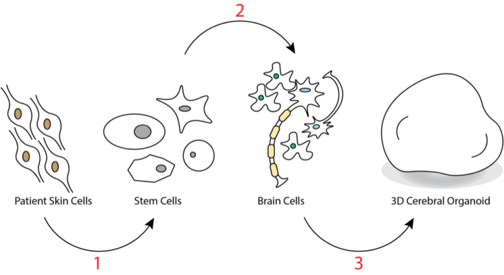
How are neuroscientists able to grow a brain in a dish, and how similar is it to an actual human brain? The process begins with stem cells. All the cells in your body have a “type”. The cells in your liver are specialized to perform the functions of the liver, for example (see a previous post about Epigenetics for more detail). The brain contains several different cell types, including the cells responsible for sending electrical signals to other cells in the brain and the rest of the body – neurons. Stem cells are cells that don’t yet have a “type” and could theoretically become any type of cell depending on the molecules instructing it. Cells in adults already have a type, but scientists have figured out how to create stem cells from programmed adult cells. They do this by taking skin cells, for example, and reprogramming them to become stem cells (see Figure 1). The stem cells can then be reprogrammed again, to turn them into neurons1. Decades of research on the molecules that determine a cell’s type are put to use growing organoids. After instructing the formation of many different types of cells that live in the brain and allowing the cells to grow in 3-dimensional space, the cells begin to mature in a manner similar to a developing human brain- forming distinct layers and even lines of communication between layers. The result is a mass of brain cells that resembles at least part of a human brain! So, to the extent that this mass functions like a human brain, how can it help us understand and treat diseases of the brain?
Modeling disease in a cerebral organoid
Growing brain cells from human tissue-derived stem cells is an extremely powerful tool because scientists can use tissue from patients with neurological disorders and compare them to tissue from healthy controls, or patients without neurological disorders. Alternatively, they can use gene-editing techniques to introduce mutations into the DNA of the organoids that are associated with disease. For example, one group of neuroscientists was able to grow a brain organoid from stem cells derived from patients with a condition called autosomal recessive primary microcephaly (MCPH)2. Humans with MCPH have much smaller brains and exhibit intellectual disability. Remarkably, organoids grown from cells taken from the patients were also smaller than control organoids. Even when grown in a dish, the organoids had qualities reminiscent of the patients’ brains. The group was able to identify a mutation in a gene that is associated with MCPH and when they introduced a functional copy of the gene into the organoid cells, they were able to cause the organoids to grow to full size. In this case, organoid technology allowed the scientists to pinpoint a critical mutation in a neurodevelopmental disease which may inform targeted treatments.
Another interesting way in which scientists use organoids is to study cancer. In one study, a group introduced mutations that caused the organoid cells to grow uncontrollably, such that they formed tumors similar to those found in certain brain cancers3. They were then able to implant these organoids in mice and show that they spread. Finally, and most importantly, they were able to screen cancer drugs to see which were able to specifically kill tumor cells while leaving healthy cells unharmed. Techniques like this may be used in the future to both test existing therapies as well as develop new ones for specific kinds of cancer and even on a case-by-case basis to treat an individual’s unique cancer. However, while these studies highlight the many possible uses of cerebral organoids for the treatment of brain disorders, there are some important limitations to consider.
Limitations and Ethical Considerations
There are several limitations of organoid technology that scientists are trying to address and that are important to keep in mind. Cerebral organoids mostly resemble cortical structures, which form the outside layer of the brain1. Many diseases, such as Parkinson’s disease and various psychiatric disorders primarily affect brain regions in deeper structures. More work is needed to understand how to direct the development of these circuits in organoids and how to connect them to other regions to model a more realistic human brain. Additionally, organoids do not have a blood supply1, so we are unable to study the interaction between the brain and circulating molecules such as hormones and nutrients. Work is currently being done to develop organoids with a blood supply to address this limitation4.
A recent article in the journal Nature brought up an interesting ethical consideration regarding cerebral organoids5. Discussing a study that found that electrical activity in developing organoids resembles that of premature babies6, the author discusses with scientists the possibility that the organoids develop consciousness. At what point does it become unethical to perform experiments and test therapies on these brain-like structures? While the experts all agree we are far from reaching this point, it is an interesting idea to keep in mind as technology advances and cerebral organoids become more and more like the complex machine inside our skulls that makes us who we are.
References:
- Amin, N.D., Pasca, S.P. Building Models of Brain Disorders with Three-Dimensional Organoids. Neuron 100, 2 389-405 (2018).
- Lancaster, M.A., et al. Cerebral organoids model human brain development and microcephaly. Nature 501, 373-379 (2013).
- Bian, S. et al. Genetically engineered cerebral organoids model brain tumor formation. Methods 15, 631–639 (2018).
- Mansour, A. A. et al. An in vivo model of functional and vascularized human brain organoids. Biotechnol. 36, 432 (2018).
- Reardon, S. Lab-grown ‘mini brains’ produce electrical patterns that resemble those of premature babies. Nature563, 453 (2018) doi: 10.1038/d41586-018-07402-0
- Trujillo, C. A. et al.Preprint at bioRxiv https://doi.org/10.1101/358622 (2018).
Image References:
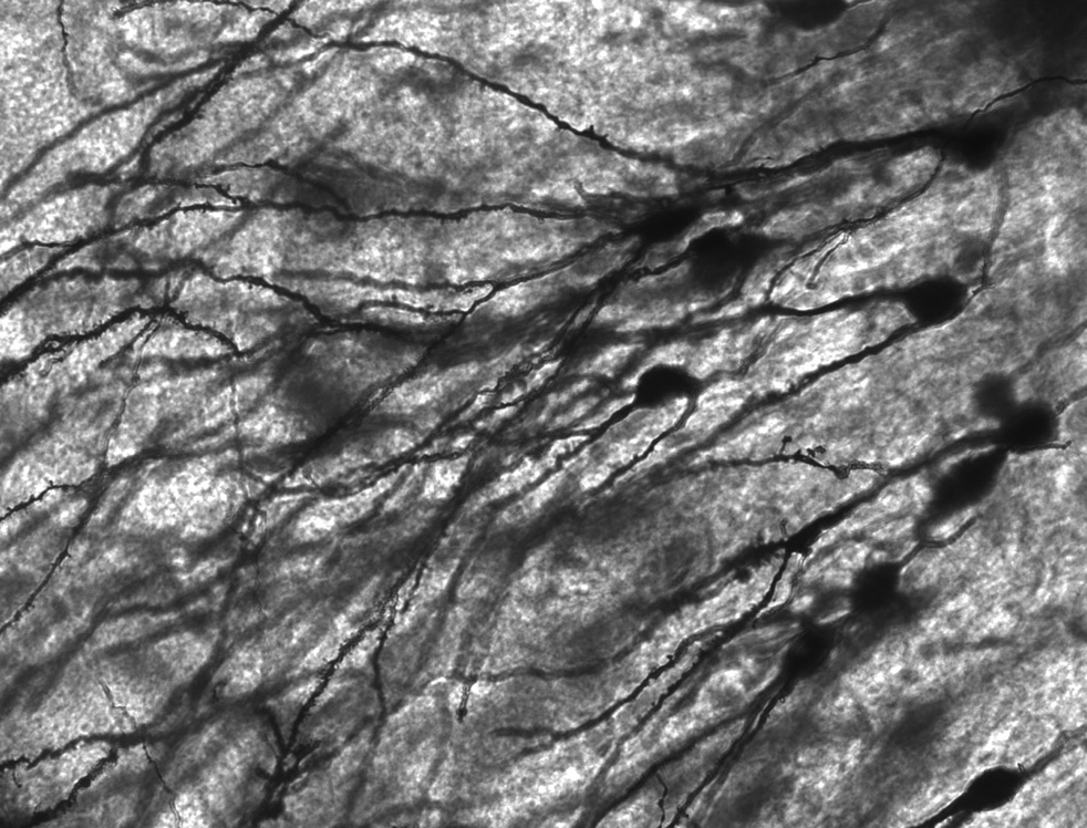
Cover Image by MethoxyRoxy via Wikimedia Commons, CC BY-SA 2.5. https://commons.wikimedia.org/wiki/File:Gyrus_Dentatus_40x.jpg
