
It’s time for our favorite annual tradition! As 2024 comes to a close, we asked our writers to share one thing that got them excited about neuroscience this year. Here’s what they said.
Lindsay Ejoh – Monkeys have names for each other?!
Did you know some monkeys have names for each other? Previously, scientists thought this kind of vocal learning only happened in humans, dolphins, and maybe elephants- but in 2024 we discovered that marmoset monkeys use special vocal patterns to name their family members. Knowing that marmosets are super social and live in close family groups, scientists set up an experiment where two marmosets from the same family were placed on either side of a physical barrier, encouraging them to ‘talk’ without seeing each other. By recording their calls, researchers discovered that the monkeys take turns speaking, almost like a conversation, and that they can identify when a unique call is directed specifically at them- sort of like hearing their names! This research gives us new insights into vocal learning and the evolution of language in the brain.
Catrina Hacker – Nobel recognition for the interplay between neuroscience and AI
Over the past few years artificial intelligence (AI) has become a common buzzword, but not everyone knows that many advances in AI were made possible by taking inspiration from the human brain. This year, the 2024 Nobel Prize in Physics recognized the mutually beneficial relationship between neuroscience and AI by awarding two scientists for their work in this space: John J. Hopfield and Geoffrey E. Hinton. Hopfield and Hinton were key players in developing artificial neural networks, which draw inspiration from the human brain and are key components of modern AI. Artificial neural networks have in turn been a rich testbed for neuroscientists to ask exciting new questions that previous tools were unable to address. It’s exciting to think how neuroscience and computer science might continue to benefit from each other in the growing field of neuroAI.
Joe Stucynski – Science drama alert! The raging debate about whether sleeping helps your brain clean itself
Earlier this year I published a post about how one of the most important things your brain does when you sleep is to clear out the molecular garbage that accumulates while you are awake. For added context, this is an established idea that neuroscientists have agreed on for over a decade. Coincidentally however, the day before that post went live, a new paper was published showing the exact opposite – that sleeping prevents molecular garbage from being cleared out, and that this process instead happens while you’re awake. Naturally, coming out and saying that the past decade of research has been dead wrong ruffled a lot of feathers, so it wasn’t long before experts in the field put together a rebuttal. While there is still more evidence that sleep aids brain cleaning than against, it’s gratifying to see the scientific process in action. Healthy debate and skepticism is vital to ensuring that scientific results are as close to the ground truth of reality as possible, no matter how popular the prevailing consensus may be.
Omer Zeliger – Sniffing out the culprit: Current research into COVID-19-related smell loss in humans and hamsters
Though it’s been five years since COVID-19 first emerged, there is still plenty we don’t know about the disease – including how it changes our noses. Losing the ability to smell is a common symptom of COVID-19 infection, and while most people recover quickly, others lose their sense of smell for months or even years. Currently, scientists think one of two things could be responsible: the virus itself is infecting and damaging neurons, or the body’s own immune system causes collateral damage when it fights off the virus. Research has been helped along by an unlikely ally: the humble hamster. These little rodents have been invaluable due to how similarly COVID-19 affects humans and hamsters, and we have them to thank for much of what we know about why the virus affects our sense of smell both short-term and long-term!
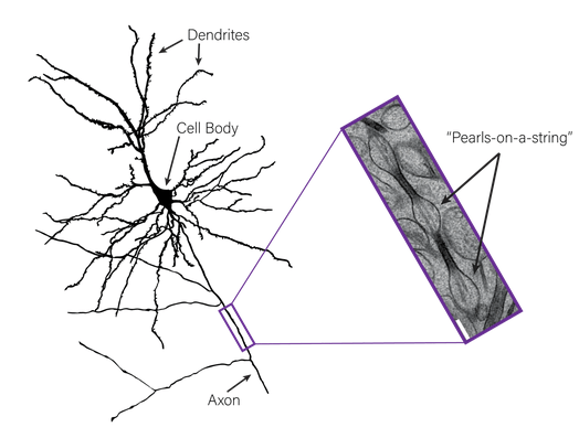
Sophie Liebergall – The cables that neurons use to communicate aren’t straight tubes, they look like a string of pearls!
Every neuron in your brain sends out a single long cable, called an axon, that it uses to transmit electrical signals to its neighbors. Historically, when neuroscientists have taken super-high-resolution pictures of axons, they looked like simple cylindrical tubes, similar to a garden hose. But before taking these high-resolution images, scientists would usually treat their brain tissue with a chemical like formaldehyde. Formaldehyde prevents the tissue from falling apart before scientists can snap a picture, but it can also distort the tissue’s structure. The Watanabe lab at Johns Hopkins University, however, are experts in a different approach: they flash freeze the brain tissue so that its structure remains much closer to how it looks inside a living animal. When the Watanabe lab flash froze some mouse brain tissue, took pictures, and zoomed in on the axons, they found that, to their surprise, they didn’t look like cylindrical tubes, but rather “pearls-on-a-string!” (Figure 1). The exact reasons for this “pearled” structure remain a mystery, however early studies hint that neurons may use the amount of “pearling” in their axon to tune the speed of the signals they send to their neighbors!
Margaret Gardner – Brain temperature isn’t just body temperature
Somewhat embarrassingly, the coolest fact I learned about this year (pun intended) is actually over 100 years old: the brain has its own temperature! Yes, I was familiar with the extremes (very high or very low temperatures) but had assumed that during daily life, your brain and body are kept at a cozy 37 degrees Celsius. However, while brain and body temperatures are strongly related, the outer areas of the brain tend to be cooler than the center (about 1 degree Celsius different in humans) and that adjacent brain regions can have different temperatures. Brain temperature also has a complicated, two-way relationship with brain activity; neural activity increases brain temperature, while even small temperature changes can change brain activity at a cellular level, impacting how likely a neuron is to activate or how many chemical messengers it takes in. So while this field may not be new, there’s still a lot for us to learn about temperature and its role in brain health and disease.
Kara McGaughey – A simple food dye can turn living skin transparent!
You’re probably well-versed at using yellow food coloring in the kitchen, but did you ever think that it might also be tremendously useful in the lab? This year, scientists uncovered a new use for tartrazine, the lemon-colored dye you’ve seen listed on almost every ingredient list as “Yellow 5.” With a deep dive into optical physics, researchers realized that tartrazine has the perfect light-absorbing and scattering properties to make living tissue appear transparent. After perfecting the dye’s formula using gel patches and slices of raw chicken, researchers gently massaged tartrazine into the skin of mice in various places. Suddenly, they could see beneath the mouse’s fuzzy surface, marveling (with the help of microscopes) at the digestive system in action, the twitch of leg muscles, or the pulse of the brain’s blood vessels. While the transparency effect lasts only 10-20 minutes, this tartrazine technology has exciting implications for visualizing the structure, activity, and function of tissues below the skin’s surface without the need for surgery. Hopefully, in the years to come this creative approach will yield new insights into the body, the brain, and how they interact to support one another.
Andrew Nguyen – Sleep heals the broken heart
We all know the importance of sleep to keep our brains and minds healthy, but scientists have found new ways in which sleep can help heal a “broken” heart. This year, a new study found that the more a patient sleeps after a heart attack the better it heals and the less inflammation there is around the heart. When the body needs to heal itself, it sends immune cells through the circulatory system to the brain to deliver a payload of proteins. Once in the brain, those proteins increase how much you want to sleep. If that sleep is disrupted, inflammation in the brain and heart both get worse and the heart is unable to heal properly. Thus, it is important for heart attack patients to sleep in order to heal their hearts. This is exciting because it shows how the body communicates to the brain via the immune system, and how the brain has evolved to regulate sleep to help the body heal properly.
Stephen Wisser – Scanning the mouse brain for genes
Just like many Americans get excited to see the latest tech Apple has developed with their annual iPhone announcement, I get excited by what new tech neuroscientists have developed every time they publish a paper. This year, that was a tool that allows a scientist to investigate what genes are active in every brain region across an entire mouse brain, called TRISCO. In a previous post, I explored the importance of technologies that allow a scientist to efficiently analyze all the active neurons in a mouse brain, an important tool to help scientists decide which brain regions to study next. But until this November, these screening techniques only told us what neurons were active after some experiment, and they didn’t say what active neurons were doing while they were active. It would be like asking a friend how their weekend was, and their response is simply good or bad. Wouldn’t it be more helpful if that friend told you why their weekend was good or bad? With this next generation of screening tools like TRISCO, we now get that extra information by seeing what genes were active and where they were active across the whole mouse brain in response to some experiment. Now that there’s a way to get more information from a brain, I wonder what we’ll learn and what secrets to diseases we’ll uncover in the new year.
Lisa Wooldridge – What’s going on “in the zone”?
You might have experienced the feeling of being “in the zone” while doing something you’re excited about or very good at. This is also known as a creative flow state. During these times, you become completely absorbed in your work and feel excited or elated, often even losing track of time. This year, our neighbors at Drexel University’s Creativity Research Lab recruited experienced Jazz guitarists and monitored their brain waves as they improvised – a technique which requires a high level of skill and creativity – to try to see what happens in the brain during a creative flow state. The researchers found that, compared to improvisations when the guitarists didn’t feel “in the zone”, high flow states were accompanied by reduced use of a set of brain areas collectively called the Default Mode Network. This “network” is active at rest – including when you’re not doing anything in particular and when you’re introspecting about your own thoughts and feelings. The Creativity Research Lab says that these findings might mean that so-called “default” brain activity can interfere with high-creativity tasks– and when it quiets down, the “task performance” part of the brain can be heard louder, unlocking the door to “the zone”.
Abby Lieberman – A virtual rodent helps us understand how the brain controls movement
This year, scientists used artificial intelligence to create a “virtual rodent” that mimics the natural movements and behaviors of a living rat! The digital rat moves in a virtual environment that accounts for real-world physics and uses a virtual brain (an artificial neural network) to control its body. Even though the scientists only specified that the virtual rodent should move like a rat (and not how to accomplish that), the artificial neural network also developed activation patterns that mimic real neural activity in biological rat brains. This exciting new approach is one way researchers might be able to use fewer live animals to study the relationship between brain activity and movement.
Jafar Bhatti – Knowing your neighbors: The first complete roadmap of an adult (fly) brain
In October of this year, after 8 long years and a major collaboration including over a dozen labs and hundreds of citizen scientists, the first complete neuronal wiring diagram of an adult fruit fly brain was published by a group called FlyWire. Although the average adult fly brain is only about the size of a poppy seed, the group found that even the brain of a tiny fly consists of nearly 140,000 neurons and more than 50 million connections (also known as synapses) between those neurons. While this might seem small in comparison to the billions of neurons and trillions of synapses in a human brain, this advancement has the potential to demystify questions related to how the organization of nervous systems enables complex behaviors such as courtship/mating, grooming, and aggression – all of which have been observed in the fly. Finally, the development of this wiring map is a notable improvement from previous maps of the worm, which only contain 302 neurons, and the larval fly, which only contain 3016 neurons. Collectively, this work is inspiring other groups to work towards a full map of the mammalian nervous system – a feat that seemed unthinkable just 10 years ago.
Nita Rome – Pregnancy Changes Brain Structure
Pregnancy has a huge impact on a person’s body, but what happens to the brain of the pregnant person during this time? To help answer this question, Dr. Elizabeth Chrastil at the University of California, Irvine had her brain imaged 26 times over the course of nearly 3 years, starting three weeks before conception, continuing throughout her pregnancy, and ending 2 years after the birth of her child. This is not the first study to collect brain imaging from pregnant people, but it is the first to repeatedly scan the same person across an entire pregnancy. The study found structural changes in the brain that became more pronounced as the pregnancy continued, with some components decreasing in volume, and other components increasing in stability. Many of these changes were temporary and returned to their pre-pregnancy baseline after the pregnancy ended, while other changes lasted for several years. While this study only includes data from one person, it marks the start of the Maternal Brain Project, an effort to repeat the study with many more people, and hopefully gain a better understanding of how the brain typically changes during pregnancy.
Emma Noel – Brain blend: one step toward personalized medicine
Brain organoids are 3D structures that model fetal brain development. To make organoids, we would normally start with stem cells taken from one patient. Recent advances have produced brain organoids generated from many individuals, termed chimeroids. Chimeroids are an incredible advancement in cell technology, because they can help illuminate cellular differences between individuals. For example, scientists treated the chimeroids with specific neurotoxic drugs and found that cells from one individual died when put in contact with ethanol (one type of neurotoxic drug), but not cells from other individuals. It will be exciting to see how chimeroids may help scientist study multiple individuals at once to produce more robust treatments.
Emma Fischer – Blood tests: Helping doctors precisely (and cost-effectively) diagnose Alzheimer’s Disease
Alzheimer’s Disease is the most common form of dementia and often leads to difficulties with a person’s memory and functioning. In a research lab setting, scientists have access to more tests to diagnose Alzheimer’s Disease than most physicians due to high cost and low availability. This year, researchers developed a highly-accurate blood test to diagnose Alzheimer’s that could be easily and readily available to physicians for patients outside of clinical trials. The blood-based test analyzes levels of a protein that has very high levels in people with Alzheimer’s Disease, called plasma phosphorylated tau 217 (p-tau217). Accurately diagnosing people with Alzheimer’s Disease is important in order to make sure people are getting necessary support or treatment for their symptoms. This test is a cheaper and more accessible tool that, when used by clinicians, could help diagnose people with more certainty and qualify people for treatments as they become more available next year!
Carris Borland– Microglia rescue neurons by connecting via tunneling nanotubes
Neurons are special cells in the brain that help to send and receive messages from all over the body. Two proteins found inside neurons that are essential to this process are tau and alpha-synuclein. Sometimes, these proteins can abnormally pile up and form tangles, making the neuron unhappy. Recently, a group of scientists showed that a type of brain cell that fights infection, called microglia, uses thin projections that connect to and remove the bad forms of tau and alpha-synuclein from the neuron.The microglia can then use these thin projections to deliver things to the sick cell that will help them heal.
Victoria Subritzky Katz – Neuralink’s Progress in 2024: Telekinesis on the horizon?
Would you like to control the virtual world with the power of your mind? Neuralink, like other neuroscience technology companies, is developing devices that make this possible—a brain-computer interface that uses brain activity to control a computer cursor. In 2024, they successfully implanted these devices in two individuals with quadriplegia (paralysis from the neck down). This technology has enabled them to browse the internet, play video games, and even design 3D objects with their thoughts—similar to what previous technologies allowed, but with higher speed and accuracy. Unlike existing technology, Neuralink’s device uses flexible threads for recording, a promising innovation that could improve performance and longevity, but early challenges have already emerged (threads retracting) and questions remain about its long-term durability, a major roadblock for the viability of brain machine interfaces. It will be interesting to follow the progress of this work, but there is a long road ahead before these devices would be widely available to clinical, let alone general, populations.

Leave a comment