May 19, 2020
Written by: Greer Prettyman
When you let your mind wander, what do you think about? Many people, when free to think about whatever they want, end up thinking about their own feelings and experiences. Studying parts of the brain that are more active during this type of free thinking compared to goal-directed activity may offer insight into introspection, something that can be hard to study naturally.
When the brain is “at rest” in an awake and alert state but not actively engaged in a goal-directed cognitive task, a specific pattern of brain activity results. This activity pattern makes up a network called the default mode network or DMN. The DMN got its name because it is comprised of regions of the brain that are active at “default” and have activity suppressed when you are engaged with a task, such as doing math problems or a language game.
Studying the default mode network aligns with a shift in the field of neuroimaging toward network neuroscience1. A network neuroscience approach includes examining the relationships between neuronal activity in different parts of the brain and the roles of these interacting networks, rather than focusing on the roles of individual brain regions or neurons.
One way to study networks in the brain is to look at their functional connectivity, or the degree to which anatomically distinct parts of the brain are activated together by the same processes. Functional connectivity is determined by measuring blood oxygen level dependent (BOLD) signal, a metric of blood flow that corresponds with neural activity. BOLD data can be recorded with an MRI while a person is resting or doing particular tasks. When activity in distinct parts of the brain increases and decreases synchronously, this correlation of activation patterns over time indicates that the regions are functionally connected with each other.
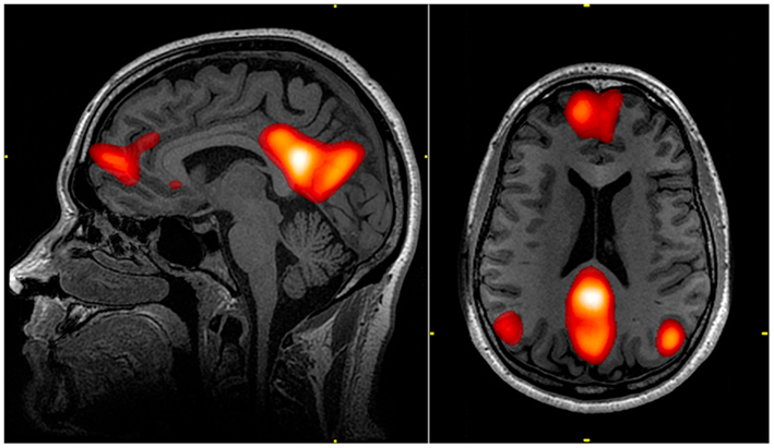
By measuring BOLD data and functional connectivity when people are at rest, researchers now know that the default mode network consists of regions including the medial prefrontal cortex, the posterior cingulate cortex, the precuneus, and bilateral parietal cortex2,3 that have high functional connectivity with each other (Figure 1). This network of regions has high activity when the brain is at rest, and activity decreases when most goal-directed tasks begin4. Other networks, in contrast, are less active at rest and become activated by tasks that require attention or cognitive work.
Why might people have areas of the brain that are more active at rest than when they are engaged? When people are not directed to think about anything in particular, they typically experience spontaneous cognition, such as wandering thoughts or daydreams. During these periods of spontaneous cognition, a majority of people report thinking about the recent past and near future5, suggesting that people gravitate toward thinking about themselves and their own lives. Activity in the DMN corresponds to this type of spontaneous cognition and thus may be necessary for self-referential thinking about ourselves and our internal states.
When a person’s attention shifts from spontaneous cognition to a task that requires external attention, such as determining which way objects on a screen are moving, activity in the DMN is suppressed6. As this task gets more challenging and requires more attention, the DMN is even more suppressed. During times when it’s important to focus on a difficult task, suppressing the DMN may be necessary to help you avoid being distracted by thoughts about your recent date or what you’ll be having for dinner.
The structural and functional connectivity of the brain change as the brain develops. Typically the DMN strengthens from childhood to adulthood and then begins to weaken during aging7. Altered development of the DMN has been observed in a variety of conditions and may offer insight into specific symptoms. For example, children with autism spectrum disorder (ASD) have reduced strength of connectivity in the DMN compared to children without ASD8. This reduced development of the DMN is thought to be related to specific symptoms of ASD such as impairment in theory of mind, or thinking about the internal states of others.
At the other end of the lifespan, accelerated weakening of the DMN during aging is associated with neurodegenerative disorders. Compared to healthy elderly people, patients with Alzheimer’s disease had disrupted activity in the DMN9. The DMN is often associated with activity in the hippocampus, a region involved in episodic memory. Reduced connectivity between DMN and hippocampus in Alzheimer’s disease may be related to problems recalling the past.
Researchers still have a lot to learn about how the brain’s networks including the DMN change over the lifespan and relate to cognitive and behavioral processes in healthy and unhealthy brains. Understanding how we think when our when our minds are free to wander remains a challenge in neuroscience research. The role of the DMN in self-referential thinking suggests that this network may be related to our representations of our consciousness. While a role for DMN in self-representation may seem uniquely human, the DMN has also been observed in monkeys, cats, and rats4. Further investigation into the role of the DMN may offer insight into how we think about ourselves.
References:
- Bassett, D. S., & Sporns, O. (2017). Network neuroscience. Nature Neuroscience. Nature Publishing Group.
- Raichle, M. E., MacLeod, A. M., Snyder, A. Z., Powers, W. J., Gusnard, D. A., & Shulman, G. L. (2001). A default mode of brain function. Proceedings of the National Academy of Sciences of the United States of America, 98(2), 676–682.
- Graner, J., Oakes, T. R., French, L. M., & Riedy, G. (2013). Functional MRI in the investigation of blast-related traumatic brain injury. Frontiers in Neurology, 4 MAR, 16.
- Raichle, M. E. (2015). The Brain’s Default Mode Network. Rev. Neurosci. 38:433–47
- Andrews-Hanna, J. R., Reidler, J. S., Huang, C., & Buckner, R. L. (2010). Evidence for the default network’s role in spontaneous cognition. Journal of Neurophysiology, 104(1), 322–335.
- Singh, K. D., & Fawcett, I. P. (2008). Transient and linearly graded deactivation of the human default-mode network by a visual detection task. NeuroImage, 41(1), 100–112.
- Mak, L. E., Minuzzi, L., MacQueen, G., Hall, G., Kennedy, S. H., & Milev, R. (2017, February 1). The Default Mode Network in Healthy Individuals: A Systematic Review and Meta-Analysis. Brain Connectivity. Mary Ann Liebert Inc.
- Washington, S. D., Gordon, E. M., Brar, J., Warburton, S., Sawyer, A. T., Wolfe, A., … Vanmeter, J. W. (2014). Dysmaturation of the default mode network in autism. Human Brain Mapping, 35(4), 1284–1296.
- Greicius, M. D., Srivastava, G., Reiss, A. L., & Menon, V. (2004). Default-mode network activity distinguishes Alzheimer’s disease from healthy aging: Evidence from functional MRI. Proceedings of the National Academy of Sciences of the United States of America, 101(13), 4637–4642.
Images
Cover Image via by Gerd Altmann from Pixabay https://pixabay.com/illustrations/artificial-intelligence-brain-think-3685928/
Figure 1 from John Graner via Wikimedia Commons, public domain https://commons.wikimedia.org/wiki/File:Default_mode_network-WRNMMC.jpg

I just knew that cats have a DMN.
LikeLike