
August 17, 2021
Written by: Marissa Maroni
According to the CDC, approximately 5.8 million Americans have Alzheimer’s disease (AD). AD is a neurodegenerative disorder characterized by the accumulation of certain proteins in the brain. One such protein is called beta-amyloid, which forms plaques outside of cells. Another is tau, which accumulates inside cells1,2. The symptoms of AD include difficulty remembering, depression, impaired communication, and confusion among many others2. With the FDA recently granting accelerated approval of a new drug called Aduhelm, the outlook on this debilitating disease is beginning to seem more hopeful. However, scientists are still working to understand the underlying biology of this complicated disease.
One major outstanding question is why specific brain regions are more susceptible to neurodegeneration than others in patients with AD. Regions important for memory like the entorhinal cortex and hippocampus, are specifically vulnerable to cell death early on in disease progression3. Why is this the case? Recent work from the University of California San Francisco (UCSF) found that a protein called ApoE may play a role in region-specific vulnerability to neurodegeneration in AD4.
What is ApoE?
The protein ApoE is a lipid (the molecular building block of fat) transporter important in lipid metabolism, a process that is necessary for energy storage5. ApoE comes in three isoforms or versions; which are named ApoE2, ApoE3, and ApoE45. ApoE4 decreases the ability for neurons to communicate properly, impairs memory, and causes tau accumulation4. Interestingly, the ApoE4 gene is the most common risk factor in certain types of AD, leading scientists to hypothesize that this protein could be important in understanding the pathology of AD and how the disease might progress throughout the brain6.
Introducing the technique: single cell RNA-sequencing
Interested in testing if the protein ApoE contributes to brain-region specific vulnerability, researchers at UCSF used a technique called, single cell RNA-sequencing (scRNA-seq). Although you may think of your brain as a web of neurons, the kinds of cells within it are very diverse! For example, the population of cells found in your hippocampus differ from cells in your cerebellum, with each region containing various cell types such as excitatory/inhibitory neurons and non-neuronal cells like astrocytes. These cells are different because they produce different proteins based on which parts of their genetic code are accessed. Single cell sequencing is a powerful technique providing scientists with a snapshot of the expression (or amount) of many genes in thousands of individual cells. With this data, scientists can compare the expression of all these individual cells and use them to answer scientific questions like “how do these cells differ from each other and how are they similar?” In the current study, scientists used this technique to see if their protein of interest, ApoE, impacts the function of individual cells differently. If so, could this difference explain why certain neurons die first in AD?
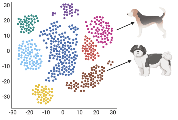
It can be hard to visualize what exactly scRNA-seq looks like so let’s take a step back and use a fun example. Imagine brain cells as a large group of dogs containing many different breeds. You, the scientist, don’t know anything about these dogs’ breeds. Picture scRNA-seq as a dog breed expert that can gather a list of characteristics about each dog (like whether they bark or howl). Using this list, you can then assign breeds based on those listed characteristics. To visualize this data, you use a computer program that can take your list of dog characteristics, group each dog based on their similarity, and label them based on specific markers. Figure 1 shows an example of scRNA-seq data based on our example. In this case, the computer program determined that the pink cluster is the “beagle” group based on their shared howling feature. Overall, scRNA-seq is a tool that can take a data set with lots of unknowns and give you clues as to the relationship between groups of cells.
What did the researchers find?
ApoE contributes to what makes brain cells different and correlates with immune pathways in mice and humans
Now that we understand the method used, let’s dive into what the scRNA-seq data told the researchers. The scientists hypothesized that differences in the expression of certain proteins might be the reason that some brain cells are more likely to degenerate in the early stages of AD. The researchers performed scRNA-seq on the hippocampus of mice that expressed a version of the protein ApoE to see if ApoE could contribute to cell-type specific vulnerability to cell death. They found that ApoE expression levels correlated to differences seen within a specific cell type. This was measured using a principal components analysis (PCA) plot. This plot is like the plot seen in figure 1 where a scientist can provide data to a computer program which can group data based on their similarity. They found that ApoE levels correlated with whatever was separating a specific hippocampal cell type from each other. Further, they found the same correlation in samples of cells from humans with mild cognitive impairment (an early stage of memory loss) and AD, suggesting that ApoE levels relate to within cell-type differences in both mice and humans.
Connecting back to our dog example, this is like if you gave all the dogs in your cohort premium dog food (this is representing expressing the ApoE protein in cells) and measured how much of this premium dog food each dog ate. Imagine that you then analyzed dog groups individually and found that in your beagle group, the amount of the premium dog food eaten correlated to some measure of within group differences (what makes one beagle different from another beagle). This experiment would tell you that the dog food (or ApoE) relates to something that is specific to some dogs (or cells) but right now, we don’t yet know what that specific thing is.
Next imagine that you investigated what factors could have made some beagles different from others based on the amount of premium dog food eaten. You found that the amount of premium dog food a beagle consumed correlated with an increase in fur softness of the beagle’s coat. From this you determined that this special diet may improve a dog’s coat. This type of investigation is what the researchers did with their scRNA-seq data in their ApoE mice and human data. They found that in hippocampal cell-types, ApoE levels correlated with genes that have a role in immune response in both mice and humans. This suggests that ApoE may contribute to cell-type specific vulnerability by affecting the brain’s immune response in the hippocampus specifically. Together, this scRNA-seq data helped scientists to identify a link in hippocampal cells between ApoE and the immune system.
ApoE peaks during early stages of cognitive decline
Next the researchers asked if ApoE levels could be tied to progression of neurodegenerative disease. The scientists collected data on ApoE expression throughout the lives of their ApoE mice. They found that ApoE levels were the highest at the onset of neuronal and behavioral deficits in mice. They found the that the data from human cells mirrored the mouse data where ApoE expression was highest during early stages of disease onset in patients with mild cognitive impairment. Together, data suggests that ApoE levels increase early on in disease onset and declines as neurodegeneration progresses.
Reducing ApoE in neurons protects against neurodegeneration
Previous research has shown that ApoE can contribute to neurodegeneration and this study provides new evidence that it may lead to selective cell death. But could removing ApoE in neurons protect against the neurodegeneration observed in this ApoE mouse model? To find out, the researchers depleted ApoE in neurons. Amazingly, they found that depleting ApoE reduced the amount of cell death. More specifically, removing ApoE prevented neuronal loss, hippocampal volume loss, and synapse loss suggesting that the cell-type specific degeneration in the hippocampus is mediated by ApoE.
What immune genes could ApoE be impacting?
The researchers previously found a strong association between ApoE levels, and proteins involved in immune response, but they wanted pinpoint specific proteins impacted. They found that an immune-related protein family called MHC-I was highly correlated with ApoE levels. MHC-I levels decreased when ApoE was depleted, and increased when ApoE was overexpressed. This confirmed that ApoE levels impact MHC-I levels. The researchers then asked if this increase in an immune response protein could be contributing to the tau aggregation seen in ApoE mice. To test this, they depleted the MHC-I protein family in neurons and found that by reducing MHC-I proteins they could protect the brain against ApoE-related pathologies such as tau aggregation.
What do all these findings mean? These data shows that ApoE may be driving selective degeneration in brain regions such as the hippocampus by increasing the expression of immune response proteins like the MHC-I protein family. Further, ApoE and MHC-I depletion protects against neurodegenerative symptoms. This research brings us one step closer to understanding AD onset making scientists better equipped to target the pathology before it spreads!
References:
1. Centers for Disease Control and Prevention. (2020). Alzheimer’s Disease and Related Dementias. Division of Population Health and National Center for Chronic Disease Prevention and Health Promotion.
2. Alzheimer’s Association. (2021) Alzheimer’s Disease Facts and Figures. Alzheimers Dement 2021;17(3).
3. NIH National Institute on Aging (NIA) (2017). What Happens to the Brain in Alzheimer’s Disease? U. S. Department of Health & Human Services.
4. Zalocusky, K. A., Najm, R., Taubes, A. L., Hao, Y., Yoon, S. Y., Koutsodendris, N., … & Huang, Y. (2021). Neuronal ApoE upregulates MHC-I expression to drive selective neurodegeneration in Alzheimer’s disease. Nature Neuroscience, 24(6), 786-798.
5. Huang, Y., & Mahley, R. W. (2014). Apolipoprotein E: structure and function in lipid metabolism, neurobiology, and Alzheimer’s diseases. Neurobiology of disease, 72, 3-12.
6. Uddin, M. S., Kabir, M. T., Al Mamun, A., Abdel-Daim, M. M., Barreto, G. E., & Ashraf, G. M. (2019). APOE and Alzheimer’s disease: evidence mounts that targeting APOE4 may combat Alzheimer’s pathogenesis. Molecular neurobiology, 56(4), 2450-2465.
Image Credits:
Figure 1 created in BioRender
Photo by Kelly Sikkema on Unsplash
