
February 11, 2020
Written by: Rebecca Somach
Here’s a riddle: What do galvanized metal, electricity and frogs have in common? If you guessed that they all have to do with one of the earliest researchers of electricity in the body, you would be correct. These concepts are all connected to Luigi Galvani. He was a scientist born in 1737 in the Papal States, which would later become Italy. His experiments in the 18th century changed our understanding of how nerves work and paved the way for electricity-based techniques in neuroscience.
We now know that the body moves because of commands that are sent to the muscles via the brain and spinal cord. However, this chain of command can’t be seen by the naked eye and also cannot be seen if an animal is cut open. When people were first studying anatomy, they were interested in understanding how muscles moved but it was not obvious how this occurred. The first theories at the time were that muscles moved because they were being inflated by fluids1 or air. These were called the ‘balloonist theories’ and were the primary ideas of how muscles moved since the earliest days of the study of anatomy and physiology by the ancient Greeks. While some experiments in the 17th centuries called these theories into question, it was Galvani’s experiments in the 18th century that began to shed light on what was actually happening.
Galvani’s most influential experiments began when he and his assistants had set up a preparation of frog legs on a table (Figure 1). At the same time, they had a machine nearby that produced electricity. It happened that one of the exposed nerves of the frogs was touched with a metal instrument while the electrical machine had been on and delivering sparks. Since this touch allowed for the conduction of electricity through the nerves, the frog legs kicked, even though the frog was definitely not alive. The observation that electricity could cause the movement of muscles fascinated Galvani and literally sparked the science of ‘bioelectricity.’ This would be the doom of the fluid- or air-based balloonist theories of muscle movement. Galvani kept up his experiments, trying different types of connections and wire materials. Furthermore, since the preparation was relatively simple, other scientists were able to replicate these findings and make their own progress towards understanding this new phenomenon. With these experiments, physiology had entered the age of electricity.

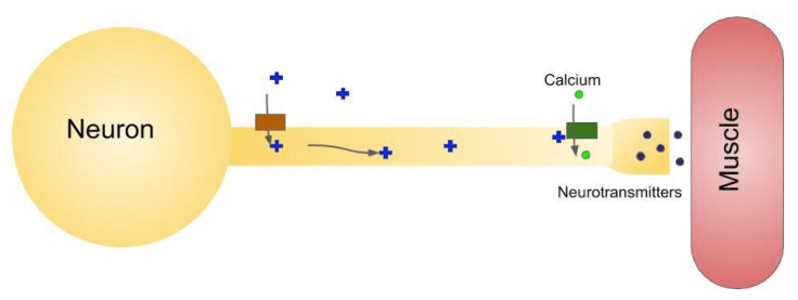
Despite the interesting experiments with electricity, Galvani did not actually know what was happening inside the body when a muscle moves. Scientists now have a much better idea of what is going on (Figure 2). The body doesn’t use electricity from an outside machine, but the neurons do use electric charge to send messages throughout the body. Neurons are designed to take advantage of the speed of electricity to make sure messages travel quickly.
In electrical devices, including the lights in a house and the device you are probably reading this article on, the device works because there is a movement of charged particles. The movement of charged particles is called an electrical current. In a neuron, charged particles also move to create something called an action potential. To get a signal from one end of the neuron to the other, the neuron is built with a set of channels that will let charged particles in and out. These channels are activated by a change in the electric field in and around the neuron. When activated, a chain reaction of channel opening begins, allowing an electrical charge to move down the axon. The axon, the long projection of the neuron, is built like a cable and is designed to let those charged particles flow in an electrical current.
When an animal wants to move a muscle, a neuron will conduct that message to the muscles in two parts. The first part of the process is the action potential2, which conducts electricity through the axon. When the electrical signal reaches the presynaptic terminal at the end of the axon, the second part of the process happens. The electrical signal is converted to a chemical one, and chemicals called neurotransmitters are released from the presynaptic terminal onto the muscle. The neurotransmitter travels from the neuron to its target muscle and causes it to move.
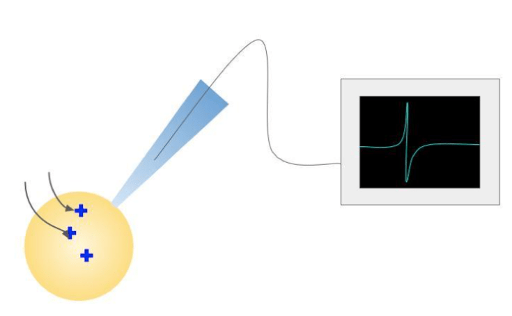
All neurons use action potentials to communicate, not just the ones that make muscles move. Many researchers have worked to understand the full picture of how neurons communicate with one another. The experiments that helped build the understanding of this system are the foundation for many other theories and experiments done in neuroscience. What are now considered ‘classic’ experiments in neuroscience have their origins in Galvani’s experiments. These include the discovery of action potentials and the Nobel Prize winning work done by Alan Hodgkin and Andrew Huxley as they looked at electricity conducted through the squid giant axon3. This work also led to the development of patch clamping (Figure 3), a technique used to examine the way that electrical currents flow into and out of cells4.
Today , the knowledge that the brain is an electrical structure is still used in a variety of research techniques and therapies. To examine brain activity during sleep as well as seizures, an electroencephalogram5, or EEG can be used. Many electrodes are placed on the outside of the scalp and a device will record how the neurons in the brain are activated together. If the electrical signals are strong enough, they can be recorded to monitor things like dreaming.
Not only do researchers and physicians use electricity to record information, but they can use electricity to influence the brain as well. Deep brain stimulation, or DBS is when a physician will implant a device in the brain that will deliver electrical signals to specific parts of the brain. DBS has been used to treat Parkinson’s disease to replace electrical stimulation that is missing due to the loss of specific neurons6. Without understanding that the brain is an organ that uses electricity as its primary method of communication, DBS, and other electrically based therapeutics would not exist. It is thanks to Galvani that the field of neuroscience continues to be the electrically charged field it is today.
References:
Piccolino M. Animal electricity and the birth of electrophysiology: the legacy of Luigi Galvani. Brain Res Bull. 1998;46(5):381–407. doi:10.1016/s0361-9230(98)00026-4
- Pearn J. A curious experiment: the paradigm switch from observation and speculation to experimentation, in the understanding of neuromuscular function and disease. Neuromuscular Disorders. 2002;12(6) 600-607. doi: 10.1016/S0960-8966(01)00310-8
- Barnett MW; Larkman PM The action potential. Pract Neurol.2007; 7 (3): 192–7. PMID 17515599
- The Nobel Prize in Physiology or Medicine 1963. NobelPrize.org. Nobel Media AB 2020. <https://www.nobelprize.org/prizes/medicine/1963/summary/>
- Segev A, Garcia-Oscos F, Kourrich S. Whole-cell Patch-clamp Recordings in Brain Slices. J Vis Exp. 2016;(112):54024. Published 2016 Jun 15. doi:10.3791/54024
- Feyissa A., Tatum W. O., Chapter 7- Adult EEG. Handbook of Clinical Neurology. 2019. 160. 103-124
- Shukla, A. W. & Okun, M.S. Surgical Treatments of Parkinson’s Disease: Patients, Targets, Devices, and Approaches. Neurotherapeutics 2014; 11, 47-59.
Image References:
Cover image by user Alexa_Fotos via Pixabay: https://pixabay.com/photos/frog-pond-animal-water-frog-4526640/
Figure 1 from Galvani, Luigi. Aloysii Galvani De viribus electricitatis in motu musculari commentarius. Bononiae: Ex Typographia Instituti Scientiarium, 1791, 10.5479/sil.324681.39088000932442. Courtesy of the Smithsonian Libraries, public domain.
Figures 2 and 3 made in Google Slides
