June 9th, 2026
Written by Meagan Olson
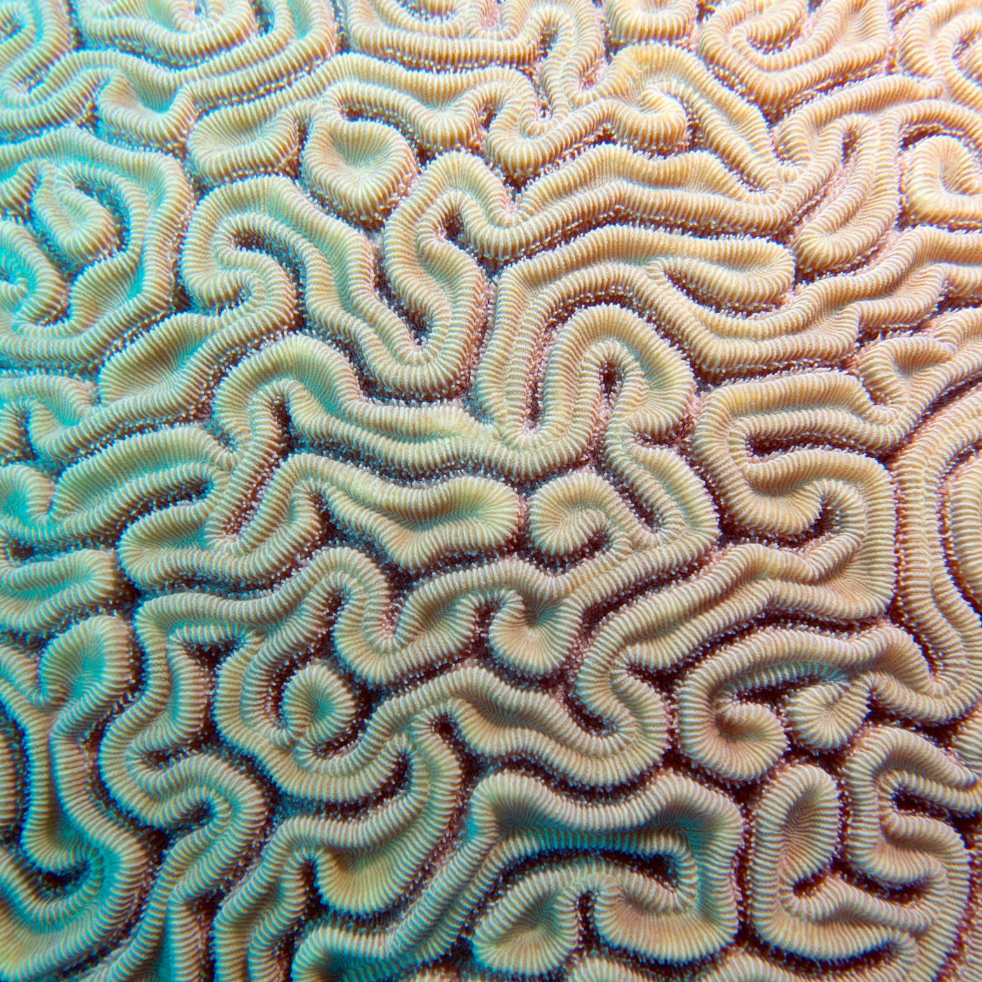
If you were to run your finger along the surface of your brain, it would feel warm, soft, and squishy. You would also notice that your brain has rounded ridges and steep grooves, giving the brain its characteristic wrinkly appearance. But why did the brain evolve this wrinkled appearance in the first place, and how do these folds form during development? In this article, we’ll explore how evolution and biology transformed the cerebral cortex into the folded structure that powers human thought and helps organize the brain into specialized functional regions.
Why did the brain become wrinkly?
The outer, wrinkly surface of the largest part of the brain is called the cerebral cortex, sometimes referred to as simply the cortex and it helps to power a variety of functions including memory, movement, sensation, language, and conscious thought1. Throughout primate evolution, increased intelligence was accompanied by dramatic expansions in the size of the cerebral cortex2. As brains became larger to support more complex thought, the cortex ran into a problem: it started to run out of space. To overcome this, the cortex began folding in upon itself as it grew, a process called gyrification3. Gyrification forms raised ridges called gyri and shallow grooves called sulci (Figure 1).

The cerebral cortex is like a thin sheet that lies on top of the rest of the brain. Although it is only a few millimeters thick4, if unfolded it would cover an area of 2.5 square feet, roughly the size of a pizza box lid4! Folding provided a way to continue expanding the cerebral cortex to this size without requiring a dramatically larger skull. By folding in on itself, the brain could pack more cortex into the limited space available within the head, creating room for additional neurons and connections. If the cortex remained smooth, accommodating more cortex would require a much larger brain.
Why do brain wrinkles matter?
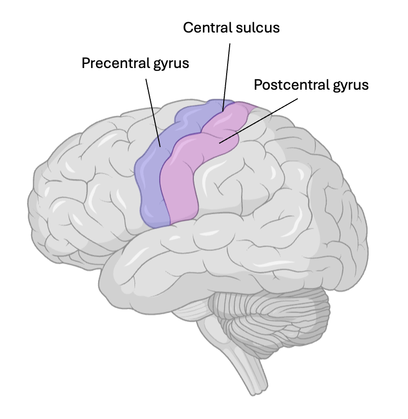
Gyrification not only solves the evolutionary problem of space, allowing more surface area to power more complex cognition, but it also helps organize this extra cortex. The major gyri and sulci form physical barriers between regions responsible for different functions, just like how the Schuylkill River separates East and West Philadelphia. For example, the central sulcus is a deep groove that runs across the top of the brain (Figure 2). On one side lies the precentral gyrus which controls body movement, and on the other side lies the postcentral gyrus, which processes sensory information like pain and touch1. Because many major folds occur in predictable locations, they also serve as important landmarks for neurosurgeons, helping them identify and navigate critical brain regions during surgical procedures!5
Wrinkles beyond the brain
The wrinkled brain is just one example of a much broader pattern in biology. Living things continuously face the challenge of limited space again and again as molecules, cells, organs, and organisms have sought to expand. Across biological scales, evolution has continuously arrived at a similar solution: folding (Figure 3). DNA, the molecule which stores genetic information, becomes tightly wound into chromosomes so that nearly two meters of DNA can fit within the nucleus of a single cell6. This packaging became increasingly important as organisms evolved larger and more complex DNA molecules while maintaining relatively small cells. Likewise, mitochondria, the organelles responsible for producing energy for the cell, has a folded inner membrane to create additional space for the molecular machinery that generates energy6. This allowed cells to meet the substantial energy demands of complex biological processes without requiring gigantic mitochondria. Biology repeatedly uses folding to generate greater complexity and function under the constraint of limited space.

Building a wrinkly brain
Scientists are still working to fully understand how exactly gyrification works. However, the leading explanation is that the brain’s wrinkles arise from differences in growth rates between different layers of the brain. During development, the cerebral cortex grows much faster than the layers of the brain underneath it. As new neurons are produced, the cortex expands rapidly, but it remains attached to the slower-growing interior of the brain. This mismatch in growth creates mechanical forces that cause the cortex to fold, forming gyri and sulci7.
The folding is not completely random, it is controlled by genetic information in DNA. The major gyri and sulci of the brain are largely the same across individuals, but the smaller folds are unique to each person8, like a fingerprint. Studies investigated the gyrification of identical twins and found that twins had more similar gyrification patterns compared to unrelated individuals9. This indicates that gyrification is heritable and controlled by genetics. Genetic information influences where neurons are produced and how quickly they populate a specific brain region3. This creates the signature pattern of major gyri and sulci that develop earlier on and are shared across individuals8. However, the minor gyri and sulci are slightly different in each person, even within twins9. This could be due to subtle variations in development. It is as if everyone is following the same instructions to create a piece of origami. The major folds appear in roughly the same places, but small differences in the process will result in each one coming out beautifully unique.
What may seem like a simple anatomical feature is actually an example of one of biology’s favorite innovations: folding. Through evolution and development, folding allowed the cerebral cortex to expand, organize into specialized regions, and support increasingly complex thought. So the next time you’re reading a PennNeuroKnow article, you have your groovy brain to thank!
References
- Purves, D., Augustine, G. J., Fitzpatrick, D., Hall, W. C., LaMantia, A. S., Mooney, R. D., Platt, M. L., & White, L. E. (2018). Neuroscience (6th ed.). Oxford University Press.
- Roth, G., & Dicke, U. (2005). Evolution of the Brain and Intelligence. Trends in Cognitive Sciences, 9(5), 250-257. https://doi.org/10.1016/j.tics.2005.03.005
- Zilles K., Palomero-Gallagher N., & Amunts K. (2013). Development of Cortical Folding During Evolution and Ontogeny. Trends in Neurosciences, 36(5), 275-284. https://doi.org/10.1016/j.tins.2013.01.006
- Chudler, E. H. (n.d.). Neuroscience for kids: Brain facts and figures. University of Washington. Retrieved June 8, 2026, from https://faculty.washington.edu/chudler/facts.html
- Tomaiuolo, F., et al. (2022). Sulci and gyri are topological cerebral landmarks in individual subjects: A proof-of-concept study. Brain Sciences, 12(8), 1005. https://doi.org/10.1111/ejn.15668
- Alberts, B., Johnson, A., Lewis, J., Morgan, D., Raff, M., Roberts, K., & Walter, P. (2022). Molecular Biology of the Cell (7th ed.). W. W. Norton & Company.
- Tallinen, T., Chung, J. Y., Rousseau, F., Girard, N., Lefèvre, J., & Mahadevan, L. (2016). On the growth and form of cortical convolutions. Nature Physics, 12(6), 588–593. https://doi.org/10.1038/nphys3632
- Borrell, V. (2018). How Cells Fold the Cerebral Cortex. Journal of Neuroscience, 38(4), 776-783. https://doi.org/10.1523/JNEUROSCI.1106-17.2017
- Schmitt, J. E., Liu, S., Neale, M. C., et al. (2021). Heritability of cortical folding: Evidence from the Human Connectome Project. Cerebral Cortex, 31(1), 702–715. https://doi.org/10.1093/cercor/bhaa254
ChatGPT version 3.5 was used to help with rewording some sentences.
Cover photo by Tam Warner Minton from Flickr.
Figure 1 made by Meagan Olson in BioRender and PowerPoint and adapted from Jmarchn on Wikimedia Commons.
Figure 2 made by Meagan Olson in BioRender and PowerPoint. Left image adapted from Thomas Splettstoesser on Wikimedia Commons.
Figure 3 made by Meagan Olson using BioRender and PowerPoint.

Leave a comment