November 3, 2020
Written by: Greer Prettyman
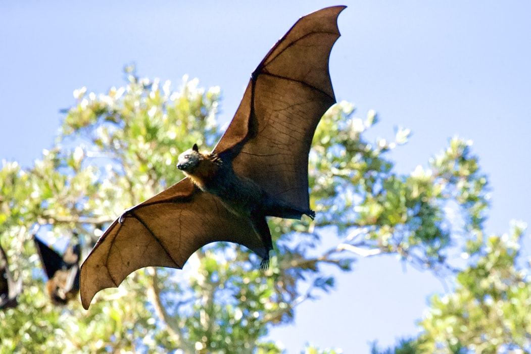
Crash. Your head automatically turns to the left so you can see your cat sitting guiltily next to a toppled pile of books. Sound familiar? The brain is really good at determining where sounds in our environment are coming from. This skill is highly advantageous for humans and animals to quickly locate the source of sounds that may signal threats, danger, and predators. For some animals, like bats, localizing sounds is also particularly important for navigating and finding food. The ears and the brain have an efficient system to locate sounds so we’re not often left wondering “where is that sound coming from?”
Sense of hearing
Sound travels through the air in the form of waves, or variations in pressure. After these sound waves enter our ear, they are translated into neural impulses that are sent to the brain that result in our sense of hearing.
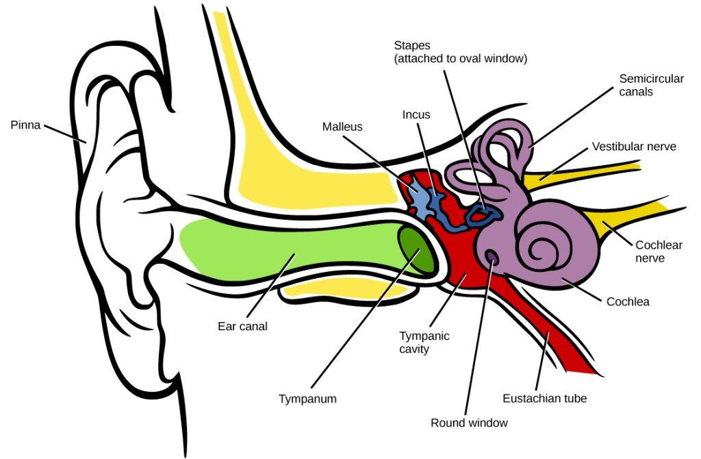
When a sound wave reaches the ear, it first vibrates the eardrum, also called the tympanum, a thin membrane separating the outer portion of the ear from the inner portion (Figure 1). In the middle ear, three small bones called the malleus, incus, and stapes help to amplify the vibrations from the eardrum1. The inner ear is full of fluid, so once the sound waves pass through the middle ear, they are no longer traveling through air, but through liquid. An organ called the cochlea gathers information from these waves in the liquid. A membrane in the cochlea called the basilar membrane has regions that are sensitive to different sound frequencies. A high frequency sound will cause one end of the basilar membrane to vibrate, while a lower frequency sound will vibrate a different region on the membrane1.
Next, the fluid waves must be converted to neural signals that can send information to the brain. Part of the cochlea called the organ of Corti is responsible for translating the sound waves across the basilar membrane into electrical impulses1. The organ of Corti is covered in small receptors called hair cells that are sensitive to tiny movements. Therefore, when sound waves moving through the liquid of the cochlea cause the hair cells to move and lengthen, they allow ions to flow, creating an electrical signal. These signals are then sent to primary auditory neurons, which form the auditory nerve. Now it’s time for the signal to travel into the brain.
The auditory neurons first enter the cochlear nucleus which is located in the brainstem, where the brain connects to the spinal cord. The auditory signal travels through several regions of the brainstem before it is sent to the thalamus, a relay center for sensory information in the brain. From there, the signal is sent to the primary auditory cortex, the part of the brain that is responsible for determining what we are hearing.
Sound localization
How does the brain determine where a sound came from? The short answer is by comparing how the sound waves hit each ear. Picture the sound waves that come from the crashing pile of books on your left side. The sound waves will enter your left ear earlier and with greater intensity than they will reach your right ear, since they have to travel a longer distance to the further ear.
The brain makes quick calculations about the timing and intensity difference between the two ears to determine where the sound must have come from. These are referred to as the interaural (between ears) time differences (ITD) and the interaural level difference (ILD). Our ears are able to detect ITDs of just a few microseconds and ILDs of a single decibel2.
These interaural calculations occur primarily in several small structures within the brainstem. Bushy cells, a particular type of neurons, in the ventral cochlear nucleus receive information from the auditory nerve2. They then send signals carrying information about timing and intensity of sounds to the superior olivary complex. Within the superior olivary complex, two separate nuclei, the medial superior olive (MSO) and the lateral superior olive (LSO), respond differently to sounds from each of the two ears.In the LSO, signals about sound intensity from the ipsilateral (same side) ear activate specific neurons, while signals from the contralateral (opposite side) ear inhibit these same neurons2. Having these two inputs on the same neurons allows them to perform a comparison: if the cell is more activated, the sound is stronger on the ipsilateral side and if the cell is inhibited the sound is stronger on the contralateral side (Figure 2). In the MSO, a similar process occurs, but the MSO is more sensitive to the difference in the timing of the sound between the ears1. Once the comparison from the inputs is completed in the MSO and LSO, the signals about the location of the sound can be transmitted to other parts of the brain, including motor areas so your head and eyes can turn toward the source of the noise.

Of course, our brains are always incorporating information from all of our senses, so we do not rely only on ITD and ILD for sound localization. Visual cues can also aid in sound localization3. For example, if you are trying to determine who is speaking in a crowd, looking at whose mouth is moving will help you to accurately locate the sound.
The bat
Humans typically only need to precisely locate whether a sound is coming from their left or their right. Bats, however, also need to account for the space above and below them while they are flying and when they are using echolocation. Bats are able to complete these impressive feats using three-dimensional sound localization.
Just like we do, bats rely on ITD and ILD to compare time and intensity differences between their two ears4. They have neurons in the MSO and LSO that do very similar calculations to those in humans in order to locate sounds to their left and right. However, bats can also move their ears much more than we can and use this ear movement to gather information based on how sounds are entering the ear canal. Bats can use only one ear to determine the position of a sound above or below them to a much finer degree than we can. By measuring frequency of spectral notches, or specific features of the sound waveform, bats are able to identify the elevation of the sound’s source5. Neurons in the dorsal division of the cochlear nucleus (DCN) respond to these spectral cues2.
Bats also use echolocation to locate objects in space using sound echoes. When echolocating, bats send out a sonar vocalization and compare the time of sound output from the time of the echo to estimate the distance between themselves and the object. A bat’s basilar membrane in the cochlea is particularly sensitive to frequencies in the range of the vocalizations they produce— this helps them to detect their own echoes with high accuracy4.
Bats use a combination of all of these cues to build a 3-D map of the sounds around them. The superior colliculus, another region in the brainstem, integrates all of the information to create this advanced representation of 3-D space. Although humans don’t have the same capabilities for 3-D localization, if you’ve ever played a game of Marco Polo and tried to find your friends in a swimming pool, you have relied on your brain’s ability to localize sounds.
References:
- Pickles, J. O. (2015). Auditory pathways: Anatomy and physiology. In Handbook of Clinical Neurology (Vol. 129, pp. 3–25). Elsevier B.V.
- Grothe, B., Pecka, M., & McAlpine, D. (2010, July). Mechanisms of sound localization in mammals. Physiological Reviews. American Physiological Society Bethesda, MD.
- Bolognini, N., Leo, F., Passamonti, C., Stein, B. E., & Làdavas, E. (2007). Multisensory-mediated auditory localization. Perception, 36(10), 1477–1485.
- Wohlgemuth, M. J., Luo, J., & Moss, C. F. (2016, December 1). Three-dimensional auditory localization in the echolocating bat. Current Opinion in Neurobiology. Elsevier
- Aytekin, M., Grassi, E., Sahota, M., & Moss, C. F. (2004). The bat head-related transfer function reveals binaural cues for sound localization in azimuth and elevation. The Journal of the Acoustical Society of America, 116, 3594–3605.
Images:
Cover Image from Unsplash user James Wainscoat
Figure 1 from Wikimedia Commons, Creative Commons Attribution 4.0 International license
Figure 2 created with BioRender

Great posts. 🙏
LikeLike