June 10, 2019
Written by: Nitsan Goldstein
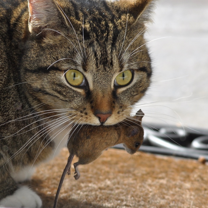
From lions hunting gazelle to your dog hunting a spider, predatory behavior in mammals is remarkably consistent. While individual hunting rituals certainly vary across species, predatory behavior is highly conserved due to its absolutely essential role in survival. Neuroscientists have been interested in the neural circuits underlying this survival behavior for years. Recent technological advances, however, have allowed several groups to use a rodent model of predatory behavior to identify specific neuron types that are critical in different aspects of hunting. In this post we’ll discuss one such study in detail. But first, we’ll venture back to the 1970s and take a brief look at the work that paved the way for this recent discovery.
Neuroscientists today are armed with unprecedented tools to manipulate and measure the activity of precise neural circuits in the brains of model organisms like mice. Decades ago, however, identifying regions of the brain associated with certain behaviors relied heavily on electrical stimulation. Electrodes would be placed into the brains of model organisms (often cats) and current would be applied to brain regions while the activity of the animal was monitored. Using these techniques, scientists were able to identify several regions that seemed to promote attack behavior. Among these were the lateral hypothalamus and periaqueductal grey (PAG)1. At the time, the conclusions that could be made were limited: these two regions are likely to somehow be involved in hunting and attack behavior. In 2018, a group at the School of Life Sciences in Beijing, China expanded upon such classical studies by using modern techniques to uncover a circuit from the lateral hypothalamus to the PAG that is sufficient to drive predatory behavior2.
The scientists thought that neurons originating in the lateral hypothalamus that receive information from brain regions that process sensory information and energy status could send axon projections to the PAG to regulate predatory behavior. To examine this idea, they trained mice to attack crickets that were placed with them in an arena. They then used a technique called calcium imaging to measure the activity of neurons that project from the lateral hypothalamus to the PAG while mice hunted the crickets. The researchers found that the activity of these neurons increased only when mice were hunting the crickets. Next, they used a technique called optogenetics to specifically activate the lateral hypothalamus projections to the PAG. They performed this experiment in mice that were not hungry or trained to attack the crickets, and therefore showed no predatory behavior at baseline. Remarkably, when they activated the neurons, the mice attacked the crickets over 90% of the time! The scientists then wondered if they could identify what kind of neurons projected to the PAG, and whether there might be other behaviors controlled by this circuit as well.
Analysis of brain tissue revealed that most of the neurons projecting from the lateral hypothalamus to the PAG were inhibitory, meaning they prevent other neurons from firing. Therefore, the scientists repeated the experiments, but this time they only recorded from and stimulated the inhibitory neurons projecting to the PAG. They found that the results were exactly the same, suggesting that the inhibitory projection to the PAG was responsible for the effect they observed previously. While analyzing the brains, however, they also found a smaller but significant excitatory projection from the lateral hypothalamus to the PAG that was distinct from the inhibitory projection. They wondered if manipulating these neurons might have a different effect. Incredibly, they found that the activity of these neurons was highest when the mouse was running away from an artificial attacker. To test if activating these neurons could drive this behavior, the scientists then built a small robot to carry food. When hungry mice were placed in an arena with the robot, they displayed aggressive attack behaviors towards it. When the excitatory neurons in the LH projecting to the PAG were stimulated, the mice immediately turned and ran away from the robot. This led the group to conclude that the inhibitory neurons in the LH that project to the PAG drive predatory behavior while the excitatory neurons are important in evasion (see Figure 1). Proper balance of these two behaviors is essential for an animal’s survival in the wild.

Future work in animals will likely continue to dissect this “hunting circuit”. For example, what neural population in the PAG are the lateral hypothalamus neurons projecting to, and where do they themselves project? How does this circuit integrate with other regions in the brain that have been shown to also be involved in predatory behavior? Finally, can studies like these tell us anything about how our own brains work? Current imaging techniques in humans are unable to specifically evaluate the relationship between lateral hypothalamus activity and aggression, however several groups have shown that the hypothalamus as a whole is involved in aggression3. Continuing to explore the neural basis for behaviors such as predatory hunting will give us a glimpse into the how the brain controls our own most primitive behaviors.
References
- Bandler, R. J. Predatory behavior in the cat elicited by lower brain stem and hypothalamic stimulation: a comparison. Brain Behav. Evol. 14, 440-60 (1977)
- Li, Y., Zeng, J., Zhang, J., Yue, C., Zhong, W., Liu, Z., Feng, Q., Luo, M. Hypothalamic Circuits for Predation and Evasion. Neuron 97, 911-924 (2018).
- Haller, J. The Role of the Lateral Hypothalamus in Violent Intraspecific Aggression—The Glucocorticoid Deficit Hypothesis. Syst. Neurosci. 12, 26 (2018).
Images
Cover Image by Stiopa and Wikimedia Commons CC 4.0 https://commons.wikimedia.org/wiki/File:Kot_z_mysz%C4%85.jpg
Figure 1 made using BioRender

Leave a comment