November 9, 2021
Written by: Greer Prettyman
Picture this: you’re back in your hometown for the holidays and go to the grocery store. As you turn into the dairy aisle, you catch sight of someone else searching for oat milk. You scan their face, which looks vaguely familiar. Are they an old friend from school, or just a stranger? After glancing furtively and searching your memories, you place them as the kid who sat behind you in math class. This process of remembering and identifying social contacts—social memory—actually relies on a different neural process than forming other types of memories.
Although mice discriminate between friends and strangers using different strategies than we do –namely, their noses—we can still take advantage of their murine social interactions to learn about how the brain assesses familiarity. A common behavioral paradigm used to measure familiarity in mice involves comparing how a mouse acts around a familiar social partner compared to a novel social partner. By assessing the amount of time that a mouse investigates each partner, researchers can make judgements about the animal’s social memory.
Social memory, unsurprisingly, depends on the brain region most commonly associated with memory, the hippocampus. Specifically, subregions of the hippocampus called dorsal CA2 and ventral CA1 are necessary for mice to remember social partners1,2. The mechanisms by which a social interaction is turned into a social memory, however, hadn’t been well understood. Recently, two studies focused on elucidating this part of the pathway to determine how mice were able to remember their friends1,2.
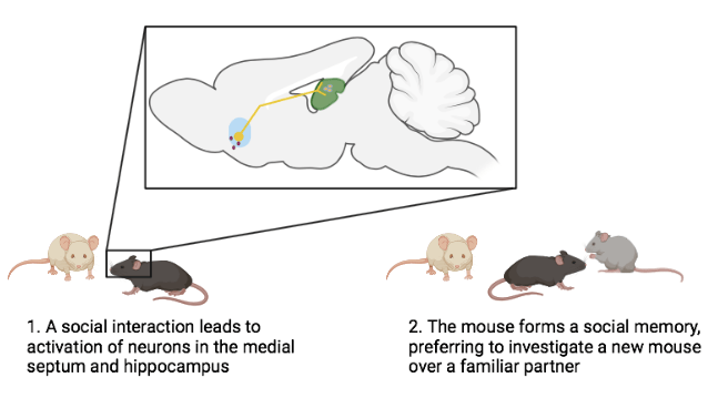
These researchers started by looking for other brain regions that send information to the hippocampus. They pinpointed two such regions, the medial septum (MS) and nucleus of the diagonal band (NDB) that were activated during social investigation and connect to the CA2 region of the hippocampus1. Activating neurons in the MS enhanced mice’s social memory; for up to 24 hours after activation, they spent more time investigating a stranger than a mouse they had previously interacted with, indicating that they remembered that familiar mouse (Fig.1). When these specific neurons that project from MS to the hippocampus were chemically inhibited, the mice could no longer form such social memories after social interactions. Non-social memory, such as remembering fear-related experiences, however, was intact. This finding indicates that this pathway is specifically required to transform a social experience into a memory.
Next, the scientists wanted to determine which specific neurotransmitters were involved in sending these social signals. The MS neurons that project to CA2 contain the transmitters acetylcholine and glutamate1. Glutamate is essential for most types of learning, and acts to increase the strength of synaptic connections, leading to memory formation. In this case, strengthening synapses between MS and CA2 hippocampal neurons during social interactions with new partners was required for social memory formation1.
The researchers now knew how the activity between the MS and hippocampal neurons drives social memory, but how do social interactions activate neurons in the MS? It turns out that another neurotransmitter, serotonin, is responsible for activating these neurons. There are serotonin receptors on the MS neurons that send glutamatergic projections to the hippocampus1. Blocking the ability of serotonin to bind to these receptors prevents the formation of social memories, while enhancing serotonin improved social memory. Interestingly, altering serotonin signaling did not change social behavior itself, indicating specific pathway for forming memories about social interactions1.
We now know a lot more about the mechanisms underlying mouse social memory, although there is still much to learn about their social identification. Given that humans have incredibly complex social interactions, the mechanisms that our brains use to determine familiarity are likely even more complex than those of mice. Unlike mice, we typically don’t rely on our friends’ scents to identify them, but we do incorporate a lot of sensory information during social interactions. Even people with prosopagnosia, who cannot visually recognize individual faces, can identify familiar people through other strategies, such as their voices. With all of this information, our brains are able to remember people we’ve known our whole lives and to form new memories of our social encounters.
References:
- Wu, X., Morishita, W., Beier, K.T. et al. (2021). 5-HT modulation of a medial septal circuit tunes social memory stability. Nature 599, 96–101
- Pimpinella, D., Mastrorilli, V., Giorgi, C., Coemans, S., Lecca, S., Lalive, A. L., Ostermann, H., Fuchs, E. C., Monyer, H., Mele, A., Cherubini, E., & Griguoli, M. (2021). Septal cholinergic input to CA2 hippocampal region controls social novelty discrimination via nicotinic receptor-mediated disinhibition. eLife, 10, e65580.
Cover image by Ingo Jakubke from Pixabay

Leave a comment