September 25, 2018
Written by: Greer Prettyman
Humans rely on feedback to learn and change behaviors. If you burn your toast one morning, tomorrow you’ll choose a lower setting. To improve at piano, you need to be able to hear when you play a wrong chord. When your friends complement your new sweater, you’ll probably wear it more often. While these may be easy changes, other behaviors, such as regulating emotions, are harder to change because feedback about your performance isn’t as easily and immediately available. But what if you could get instantaneous feedback from inside your own brain?
With new neuroimaging technologies, people can get real-time feedback about the neural activity in different parts of their brains. Using this feedback, they can then learn to alter their brain activity more effectively. Real-time fMRI neurofeedback is currently being used to target complex behaviors associated with post-traumatic stress disorder (PTSD), depression, and nicotine addiction, among others. With this new type of brain-computer feedback, people can get better at changing behavior by first learning to change their brains.
How does real-time fMRI feedback work?
Functional magnetic resonance imaging (fMRI) is a non-invasive imaging technology that uses a strong magnet to look at patterns of blood flow in the brain. The flow of oxygen-carrying blood to an area is associated with the amount of underlying neural activity, allowing researchers to indirectly measure activation of different areas of the brain over time.
During real-time neurofeedback studies, people lie down in the MRI scanner, just like they would for a clinical MRI. Researchers begin by measuring activation in a particular region of interest (ROI) in the brain. The chosen region depends on the specific behaviors that are being targeted. For example, to target fear-related behaviors in people with PTSD, activity would be measured in the amygdala, brain region associated with fear, anxiety, and emotion processing.
Scientists record signals from the region of interest and analyze it in real time to determine the level of activity. They then convert neural activation levels to a visual format that can be displayed on a screen to the person in the scanner. For example, the amount of activity in the amygdala might be visualized as bars on a thermometer that rise and fall as activity in the region increases and decreases (Figure 1).
Next, the person in the scanner uses the visual feedback to try to change their brain activity. Sometimes this happens implicitly, where the person is simply told to “decrease the bar on the thermometer” and they must try to figure out a strategy that lowers amygdala activity without being aware that that’s what they are doing. Other studies have used more explicit instructions like “try to decrease your feeling of fear.” The visual feedback about brain activation updates in real-time so people can see the effects of their efforts and learn which strategies lead to changes in activation in the region of interest.
As with any new skill, learning to control brain activity with real-time feedback requires practice. Typically, participants complete multiple scanning sessions where they work on changing their brain activity. Over time, they can learn better and better strategies for changing activity levels.
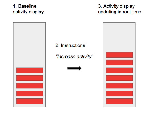
First, while a person lies in the MRI scanner, activity is measured from a specific region of the brain. Baseline activity level in the brain region is processed by a computer and displayed to the participant on a screen. Next, participants receive instructions to try to change the activity level. They may use various strategies to try to change the activity levels, such as thinking about particular things. The computer continues to process activity level and the visual display updates in real-time as brain activity changes. This allows people to visualize how brain activity changes and ultimately to learn which strategies are most effective for changing the activity level.
Many studies also look at changes in functional connectivity, or amount of communication, between different regions of the brain after a person does real-time fMRI feedback training. Disruptions in functional connectivity between various regions are associated with many psychiatric disorders. However, the amount of connectivity between two regions is not static. The connectivity in the brain is plastic, meaning the amount of communication can be strengthened and weakened depending on internal thought patterns and external factors such as the environment. Repeated practice of real-time fMRI feedback can have long term effects on connectivity, leading to changes that last long after the person leaves the scanner. For example, a recent study in veterans with PTSD found that real-time neurofeedback strengthened the functional connectivity between the supplementary motor area and the dorsal anterior cingulate cortex, a network involved in emotion monitoring and regulation. These changes in connectivity following neurofeedback training were associated with a reduction in PTSD symptoms1.
How can neurofeedback change behavior?
Numerous studies provide evidence that learning to alter brain activity with real-time fMRI feedback leads to changes in a variety of hard-to-change behaviors. In one recent study, researchers used neurofeedback to improve motivation. In this study, healthy people used fMRI feedback to regulate activity in the nucleus accumbens (NAc), a region involved in reward and motivation processing2. Participants were instructed to try to move a dial reflecting NAc activity by thinking about a positive event in the upcoming three months, a strategy that has been shown to increase activity in this region. Those people who received feedback and successfully learned to increase activity in the NAc displayed behavior consistent with higher motivation when they completed tasks to assess effort in the lab following neurofeedback training. A control group that received “sham” feedback that did not actually reflect changes in their NAc activity did not improve in motivated behavior. The ability to increase motivated behavior using neurofeedback has implications for increasing workplace productivity and healthy behaviors, as well as improving disorders that impair motivation, such as depression and schizophrenia.
Another behavior that is characteristically difficult to change is nicotine craving. Cigarette smokers often experience strong cravings for nicotine when they see smoking cues, like a picture of a cigarette. In a study out of the Medical University of South Carolina, nicotine smokers completed real-time fMRI feedback training sessions3. When instructed to “reduce craving”, these smokers were able to decrease activation in the anterior cingulate cortex, a region highly associated with cigarette craving. They also reported lower levels of craving following the neurofeedback session, suggesting that this method may be an effective way to help people to successfully quit smoking by reducing neural activity associated with craving.
These are just a few examples of ways in which neurofeedback has been used to change behaviors. Other studies have investigated real-time fMRI feedback to target behaviors associated with depression, anxiety, pain, ADHD, and even spider phobia. As a non-invasive and low-risk intervention strategy, real-time fMRI neurofeedback is a promising new way to alter neural activity and functional connectivity in the brain. fMRI neurofeedback will likely be incorporated into therapy plans as a tool to help people with psychiatric disorders change disruptive thought patterns. Neurofeedback might also help you stay motivated to work out, quit smoking, or concur your fear of spiders by learning from what’s going on inside your brain.
References:
- Misaki, M., Phillips, R., Zotev, V., Wong, C.-K., Wurfel, B. E., Krueger, F., … Bodurka, J. (2018). Real-time fMRI amygdala neurofeedback positive emotional training normalized resting-state functional connectivity in combat veterans with and without PTSD: a connectome-wide investigation. NeuroImage: Clinical, 20, 543–555.
- Li, Z., Zhang, C.-Y., Huang, J., Wang, Y., Yan, C., Li, K., … Chan, R. C. K. (2018). Improving Motivation Through Real-Time fMRI-Based Self-Regulation of the Nucleus Accumbens. Association, 32(6), 764–776.
- Canterberry, M., Hanlon, C. A., Hartwell, K. J., Li, X., Owens, M., LeMatty, T., … George, M. S. (2013). Sustained Reduction of Nicotine Craving With Real-Time Neurofeedback: Exploring the Role of Severity of Dependence. Nicotine & Tobacco Research, 15(12), 2120–2124.

Leave a comment