January 28th, 2025
Written by: Emma Noel
Scientific advances are frequently tackling the most difficult diseases, rapidly finding treatment and even cures to diseases once deemed incurable. Smallpox was largely eradicated in the U.S. by 1980 due to the creation of a vaccine1, HIV/AIDS became treatable with the discovery of a drug2, the discovery of penicillin shaped our response to bacterial infection3. Yet, cancer remains one of the hardest diseases to treat, let alone cure. Glioblastoma is an aggressive brain cancer with no current treatments, but a recent scientific advancement has brought us one step closer to finding a cure.
What is glioblastoma?
Glioblastoma is one of the most prevalent and pervasive brain cancers. The cancer primarily spreads through a young, rapidly-dividing and invasive group of cells called glioma stem cells. Glioma stem cells bud off of cancerous clumps and migrate freely in the brain, allowing the cancer to aggressively spread. Glioma stem cells are difficult to target clinically without disrupting or killing healthy brain cells, which is why glioblastoma is one of the deadliest cancers4. Despite the current poor prognosis for glioblastoma, there is still hope for the future. Many tools have been developed to study and eventually combat glioblastoma. One important tool is patient-derived brain organoids. Brain organoids are groups of cells grown from real patient cells that can be used to study brain development on a small-scale. For example, Dr. Guo Li Ming and Dr. Hongjun Song at UPenn used brain organoids to study tumor formation. In this post I’ll extend on that to discuss new innovations in brain organoid technology that could help identify treatments for glioblastoma.
What is a brain organoid?
Organoids are a clump of human cells grown in a lab that can recreate developmental steps in a dish5. For example, brain organoids can mirror small parts of human brain development, while liver organoids model liver development on a small scale. As opposed to typical cell culture which lies flat in a dish, brain organoids are 3D which makes them more similar to brain tissue. In a 3D structure like an organoid, cells grow and interact in all directions, creating layers and interactions similar to those in real brain tissue. This is different from 2D cell cultures, where cells spread out in a single flat layer and lack the complexity of real brain. Brain organoids are therefore uniquely positioned to study development. They are created by taking an individual’s blood cells and reprogramming them so that they turn into cells capable of recreating the earliest stages of neurodevelopment. The cells in a brain organoid can divide, grow, and spread, creating a structure that resembles the brain region they are modeled after6. In summary, while brain organoids can model specific features of brain development, they are not themselves considered brains.
Brain organoids are a great model for development, and recently scientists have also started to use organoids to understand how diseased human brain cells may react to treatments7. This is exciting because brain organoids can be generated from any person’s cells, making it possible to see how one individual’s “brain” responds differently than another individual to treatment. However, brain organoids have major limitations that hinder drug testing. For example, they take a long time to grow and they vary in size7. (Read more about the pros and cons of using brain organoids in this NeuroKnow article written by Sophie Liebergall).
Recently, a group of researchers in Germany took advantage of the promise of brain organoid technology to identify drugs that slow or stop glioma cell invasion of brain tissue8. To get there, they had to solve two big problems. First, they had to shorten the time it takes to make organoids. Second, they had to create a model to observe glioma stem cell invasion into the organoid.
Obstacle #1: Speeding up the organoid creation process
The researchers chose in the first place to develop brain organoids to approximate the patient’s healthy brain tissue, and see how glioma stem cells are then able to infiltrate and invade that healthy tissue. However, the researchers faced many technical challenges in developing the brain organoids needed for their drug testing. The first challenge was that brain organoids are very time consuming to make, resulting in months dedicated to creating and growing just one batch. This makes it difficult for scientists to quickly take insights from the lab to patients. Additionally, one use of brain organoids is to test how an individual with a disease responds to treatment before administering that individual the treatment. In these cases, quick results are crucial to help doctors treat the patient effectively.
In order to make organoids from an individual’s cells, scientists must first draw blood to isolate blood cells5. The cells then go through various steps to change the role they will eventually play in an organism, or their fate. You can think of the fate of a cell as similar to having a job, like some people are nurses and others are teachers, some cells work in the bloodstream, whereas others perform functions in the brain. Through this process, scientists can change the fate of the blood cells into “unfated” cells, or cells that do not have a task yet, to brain cells. From there, scientists grow the cells into a 3D brain organoid. This organoid can then be tested with drugs to see how the donor individual’s own cells react to a treatment9. Therein, scientists can figure out how a treatment works on brain cells originating from an individual, before giving that individual the treatment.
Most approaches to making organoids are extremely tedious, because each clump of cells has to be initially placed and grown in an individual compartment. Every day, the organoids have to be fed, and every week the organoids have to be transitioned to a different environment10. Given this, the researchers set out to eliminate these tedious steps and make large batches of organoids from different human cells quickly. To do so, they used a spinning machine that allows the creation of hundreds of organoids at once7. This spinning machine made it much easier to quickly grow many organoids that the scientists could then use for testing. After the development of a faster brain organoid generating system, the researchers wanted to use this system to study glioma stem cell invasion.
Obstacle #2: Measuring glioma stem cell invasion
The second main goal of the researchers was to figure out how glioma stem cells move into (or invade) the human brain7. This is an important goal, as modeling this aspect of glioblastoma would improve researchers’ ability to predict how an individual with glioblastoma may react to treatment.
Researchers studied how glioma stem cells invade brain tissue by placing a brain organoid next to the cells and tracking their movement. They found that as the glioma stem cells moved into the organoid and caused changes similar to those seen in tumors from human patients. Being able to watch how glioma stem cells invade a brain organoid is a huge advantage of using this system. This result suggests that the organoid model closely mimics the invasive behavior of glioma stem cells, providing an accurate representation of tumor progression. By tracking glioma stem cell movement into organoids, this research helps us see how cells contribute to tumor growth and spread in the brain.
Next, the researchers used this newly developed system to test over 180 potential drugs to identify which drugs could stop glioma stem cell invasion. The 180 drugs are used to treat other known diseases, and the scientists wondered if they might also be effective in treating glioblastoma. Whereas it would be difficult to test this many drugs in human patients or animal studies, the ability to make many organoids for the same patient allows the researchers to test many drugs on the organoids and then test only the most effective candidates in human patients. One of the major benefits of repurposing drugs to treat disease is that many of them have already undergone extensive and necessary safety testing. Repurposed drugs are thereby less likely to have unknown side effects and, if effective for a different disease, can quickly be moved into clinical trials and FDA approval.
Using their system, the researchers were able to identify two drugs that stopped glioma cell invasion. The candidate drugs will undergo further testing to see if they may be effective in a clinical trial for glioblastoma.
The future of glioblastoma research
The researchers have made incredible advancements in the field of glioblastoma, which could eventually be used to assess which drugs are most effective on which individuals. Moreover, they push the boundaries of organoid technology by inventing a system to standardize brain organoids derived from different individuals. Using different individuals’ tumor cells allows researchers to directly ask: how will this person’s brain cells respond to treatment, and is this treatment effective for this person? This approach could revolutionize the way we treat brain cancer, moving away from a one-size-fits-all model to a more personalized, patient-specific strategy. Ultimately, it means that we could eventually choose treatments based on how a patient’s own cells react, significantly improving treatment outcomes and minimizing unnecessary side effects. Methods like the one outlined here, the study of glioma stem cell invasion into brain organoids, may be the key to identifying new treatments for glioblastoma and possibly ending the longevity of glioblastoma as one of the most deadly cancers.
References
- CDC, About Smallpox. October 22, 2024. Cdc.gov
- National Cancer Institute. The First AIDS Drugs. https://ccr.cancer.gov/news/landmarks/article/first-aids-drugs
- American Chemical Society. Discovery and Development of Penicillin. Acs.org
- Rodriguez, Silvia M., Georgiana-Adeline Staicu, Ani-Simona Sevastre, Carina Baloi, Vasile Ciubotaru, Anica Dricu, and Ligia G. Tataranu. “Glioblastoma Stem Cells—Useful Tools in the Battle against Cancer.” International Journal of Molecular Sciences 23, no. 9 (2022). https://doi.org/10.3390/ijms23094602.
- Kim, Jihoon, Bon-Kyoung Koo, and Juergen A. Knoblich. “Human Organoids: Model Systems for Human Biology and Medicine.” Nature Reviews Molecular Cell Biology 21, no. 10 (October 1, 2020): 571–84. https://doi.org/10.1038/s41580-020-0259-3.
- Bagley, Joshua A, Daniel Reumann, Shan Bian, Julie Lévi-Strauss, and Juergen A Knoblich. “Fused Cerebral Organoids Model Interactions between Brain Regions.” Nature Methods 14, no. 7 (July 1, 2017): 743–51. https://doi.org/10.1038/nmeth.4304.
- Kim SH, Chang MY. Application of Human Brain Organoids-Opportunities and Challenges in Modeling Human Brain Development and Neurodevelopmental Diseases. Int J Mol Sci. 2023 Aug 7;24(15):12528. doi: 10.3390/ijms241512528. PMID: 37569905; PMCID: PMC10420018.
- Ramani, Anand, Giovanni Pasquini, Niklas J. Gerkau, Vaibhav Jadhav, Omkar Suhas Vinchure, Nazlican Altinisik, Hannes Windoffer, et al. “Reliability of High-Quantity Human Brain Organoids for Modeling Microcephaly, Glioma Invasion and Drug Screening.” Nature Communications 15, no. 1 (2024): 10703. https://doi.org/10.1038/s41467-024-55226-6.
- Bose S, Clevers H, Shen X. Promises and Challenges of Organoid-Guided Precision Medicine. Med. 2021 Sep 10;2(9):1011-1026. doi: 10.1016/j.medj.2021.08.005. PMID: 34617071; PMCID: PMC8492003.
- Qian, Xuyu, Yijing Su, Christopher D. Adam, Andre U. Deutschmann, Sarshan R. Pather, Ethan M. Goldberg, Kenong Su, et al. “Sliced Human Cortical Organoids for Modeling Distinct Cortical Layer Formation.” Cell Stem Cell 26, no. 5 (May 7, 2020): 766-781.e9. https://doi.org/10.1016/j.stem.2020.02.002.
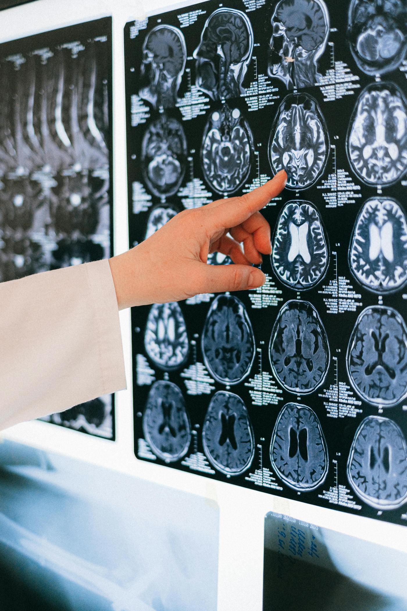
Cover Photo by Anna Shvets, Pexels.com

Leave a comment