
February 13th, 2024
Written by: Sophie Liebergall
Perhaps my most embarrassing moment as a medical student came during my second year when I was doing a clinical rotation in the emergency department. I had just seen a patient who had come in early that morning with a very deep cut on his hand. All seemed fine as I was evaluating the patient in their room, but when I went out into the hallway to retrieve the suture kit, something was amiss. I started to feel woozy. My palms suddenly glistened with sweat. The familiar walls of the emergency room began to blur before they disappeared completely. I tried to stagger towards a chair, but before I made it, I fainted onto the linoleum floor. I woke up to a flurry of doctors and nurses putting in IVs and strapping on monitoring devices. But some quick medical tests revealed that I was completely fine (even if my pride was a little bruised).
How is it possible that simply seeing an especially gnarly wound could lead to a physical response so dramatic that I completely lost consciousness? Over the last few decades, scientists have developed a solid understanding of how heart rate and blood flow change during fainting spells. But, the pathways which connect seeing something disturbing to these physical changes have remained elusive. That is, until a research group at the University of California in San Diego (UCSD) recently published an article1 which may have solved a major piece in the puzzle of fainting.
Fainting is caused by temporary decreases in blood flow to the brain
The medical term for the type of fainting that occurs due to an external trigger such as seeing blood, getting an injection, experiencing a spurt of intense pain, or standing up too fast is vasovagal syncope. As in other kinds of fainting, vasovagal syncope results in the loss of consciousness because the brain temporarily isn’t receiving enough blood flow.2
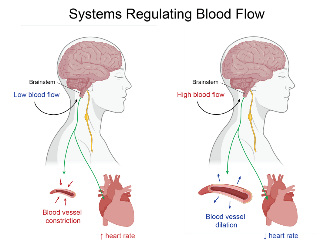
Making sure that all of the organs in the body (especially the brain) are receiving enough blood flow is incredibly important to staying alive. As such, we have evolved extremely sensitive systems to subconsciously control the delivery of blood to our organs (Figure 1). These systems first sense the amount of blood flow using special nerve cells that reside in our major blood vessels. Then, they send a report of the blood flow to a small collection of neurons in a region of the brain called the brainstem. The neurons in the brainstem then respond to the signals by changing our heart rate and constricting (narrowing) or dilating (expanding) our blood vessels.3 Speeding up the heart rate increases the speed of the blood flow; whereas constricting the blood vessels increases the pressure of the blood flow. Thus, when the brainstem neurons receive an alarm signal that the body’s organs aren’t receiving enough blood flow, they respond by telling the heart to speed up and the blood vessels to constrict. On the other hand, when the brainstem neurons receive a signal that the blood flow is too high, the neurons in the brainstem tell the heart to slow down and the blood vessels to dilate.
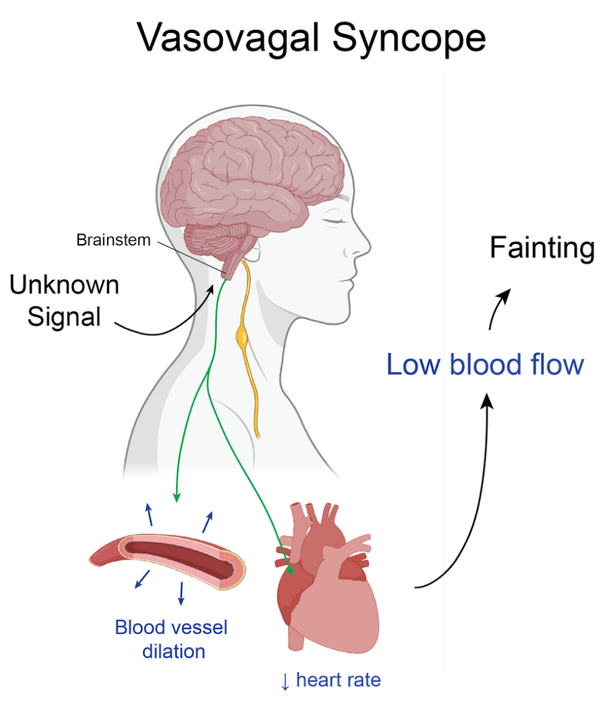
In the case of vasovagal syncope, the systems controlling blood flow malfunction. The blood flow to the organs isn’t too low. But, an external trigger (seeing blood, standing up too fast, etc.) activates some mystery sensor which causes the heart rate to mistakenly slow down and the blood vessels to dilate (Figure 2). These changes lead to decreased blood flow to the brain and loss of consciousness. But the identity of the mystery sensor sending these erroneous signals was unknown – that is until the researchers at UCSD discovered a set of neurons who seem to fit the bill!
Newly discovered neurons that sense signals in the heart can trigger fainting
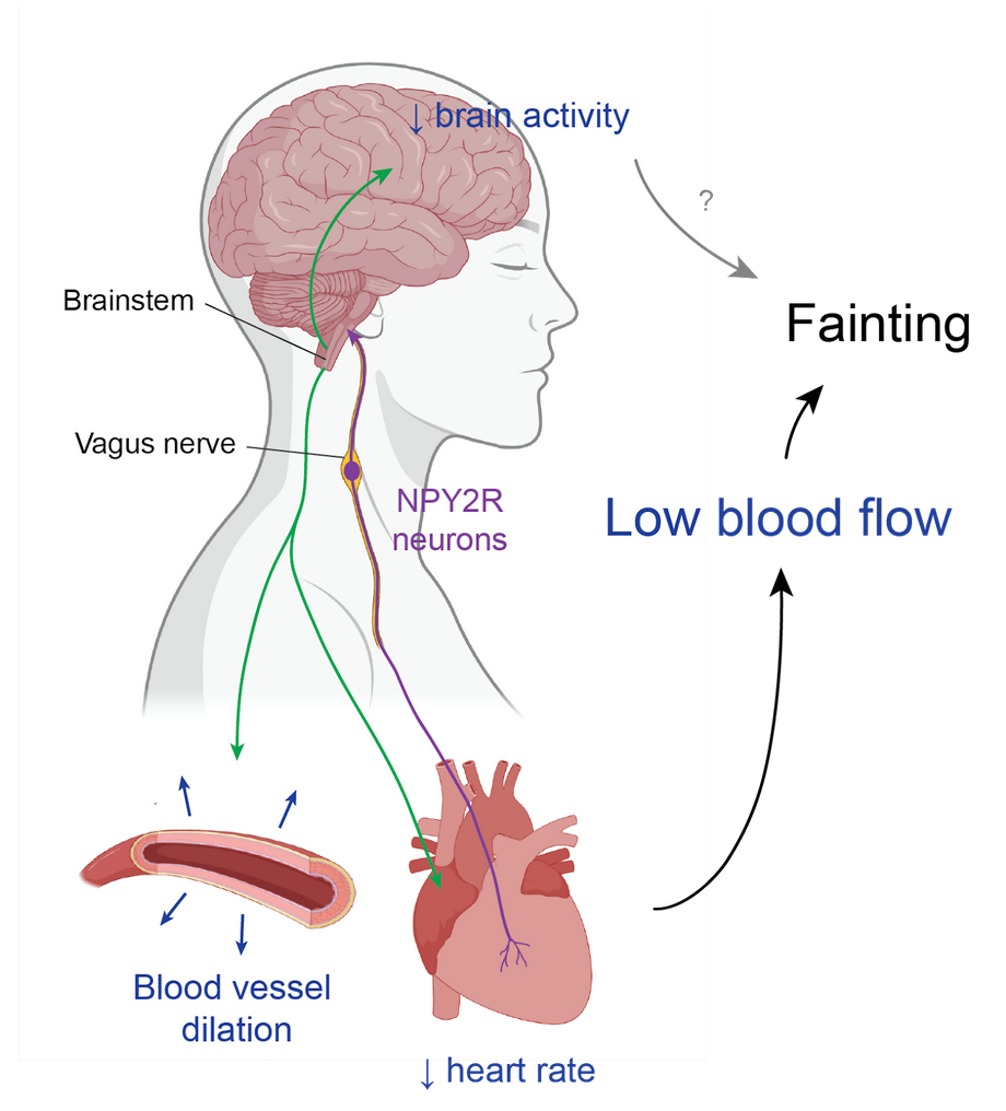
A significant portion of the neurons that sense signals important for subconscious physical properties like digestion, breathing, and heart rate travel along the vagus nerve, which is a direct wire between these organs and the brainstem. The research group at UCSD started their work by looking for previously unstudied populations of neurons in the vagus nerve. In their survey, they found a population of neurons which make copies of a gene called NPY2R. They then used a technique involving colored proteins to track where the NPY2R neurons sense information and then send their signals. This experiment showed that the NPY2R neurons sense information from the bottom part of the heart and send those signals up to the brain stem (Figure 3).
Next, the researchers wanted to figure out what these NPY2R actually do with the information that they receive from the heart. To answer this question, the researchers used a powerful technique called optogenetics, which allows them to selectively activate these neurons by shining light on them. When they activated the NPY2R neurons in mice, they were surprised to find that the mice instantly fell over and remained still before recovering and going about their business in their cage.
This response in the mice looked an awful lot like an episode of fainting! But, the researchers still had to do a number of experiments to be sure of exactly what was happening to the mice when the NPY2R neurons were stimulated. They found that the response seen in the mice very closely resembles what happens in humans when they have vasovagal syncope: their heart rates dropped, their blood vessels dilated, their eyes rolled back, there was decreased blood flow to the brain, and their brain activity showed “flattened” signals identical to fainting humans.
NPY2R neurons also directly decrease brain activity
In addition to learning that NPY2R neurons can cause vasovagal syncope, the researchers made another surprising discovery which challenges some of our classic assumptions about the mechanisms of fainting. It has been long thought that the decrease in brain activity that occurs during vasovagal syncope is a direct result of the decrease in blood flow to the brain. To test this assumption, the researchers activated the NPY2R neurons while also giving the mice a drug which ensures that the heart rate and blood pressure don’t drop. As expected, the drug prevented the drop in blood flow to the brain. But, surprisingly, the drug didn’t completely stop the mice from having vasovagal syncope. Instead it only delayed it. This suggests that some of the changes in the brain associated with vasovagal syncope may be the direct result of connections from the brainstem to the rest of the brain, rather than only secondary to the effects of decreased blood flow to the brain.
Next steps in the science of fainting
Though the researchers at UCSD have shown that activating NPY2R neurons in the vagus nerve reliably leads to fainting, there are still many unanswered questions about this newly discovered type of neuron and the general mechanisms of fainting. The researchers determined that these neurons mainly receive signals from the heart (which they then send up to the brainstem). But it is still unclear exactly what information in the heart these neurons are trying to sense, and how these neurons relate to some of the external triggers of vasovagal syncope (like seeing blood).
In addition to discovering what information the NPY2R neurons sense, scientists still need to work out the exact identities of the neurons in the brainstem that are activated by NPY2R neurons. Once they do, scientists can then understand how these brainstem neurons more directly lead to fainting.
The discovery of the NPY2R neurons hasn’t just been important in advancing our understanding of the role that the nervous system plays in fainting. It also introduces potential avenues to develop treatments for patients who are extremely prone to fainting, which can be dangerous and life-disrupting. For example, scientists could begin to develop drugs or devices which work by selectivity silencing NPY2R neurons or the brainstem neurons to whom they send their signals.
For now, though, the best treatment for fainting at the sight of blood is still exposure therapy or avoiding vasovagal triggers. And interestingly I’ve gone on to stitch up many a bloody hand without issue since that day. So what transpired on that fateful morning in the emergency department may still remain one of the great mysteries of the brain…
References
- Lovelace, Jonathan W., Jingrui Ma, Saurabh Yadav, Karishma Chhabria, Hanbing Shen, Zhengyuan Pang, Tianbo Qi, et al. “Vagal Sensory Neurons Mediate the Bezold–Jarisch Reflex and Induce Syncope.” Nature 623, no. 7986 (November 2023): 387–96. https://doi.org/10.1038/s41586-023-06680-7.
- Adkisson, Wayne O., and David G. Benditt. “Pathophysiology of Reflex Syncope: A Review.” Journal of Cardiovascular Electrophysiology 28, no. 9 (September 1, 2017): 1088–97. https://doi.org/10.1111/jce.13266.
- Eckberg, Dwain L., and Peter Sleight. Human Baroreflexes in Health and Disease. Oxford University Press, 1992. https://doi.org/10.1093/oso/9780198576938.001.0001.
Cover photo: “A lady fainting after bloodletting.” Oil painting after Eglon Hendrick van der Neer. Neer, Eglon van der, 1635 or 1636-1703. Wellcome collection 44763i.
Figures 1-3 made by Sophie Liebergall using Biorender and Adobe Illustrator.

Leave a comment