
November 28, 2023
Written by: Julia Riley
Parkinson’s disease is a multi-faceted illness that affects the way people move, think, sleep, and even feel1–3. It’s also incredibly common. The number of people in the US currently living with Parkinson’s is about 20% more than the entire population of San Francisco3. We have succeeded in developing several treatments for Parkinson’s that help make symptoms more manageable for patients and their families. However, although Parkinson’s is common and diagnosis rates are only projected to increase in coming years, we have yet to develop a true cure for the disorder. You may be wondering- if Parkinson’s is so common, why has a cure escaped us for so long?
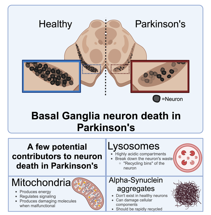
Usually, we have to know what causes a disease in order to cure it. This is because most cures are designed to stop an illness at its source. We have discovered that the symptoms observed in Parkinson’s stem from the death of neurons in the basal ganglia, a part of the brain that controls movement. Specifically, this brain region helps you smoothly start and stop movements. Like most regions in the brain, neurons in the basal ganglia are special in that they are irreplicable- when neurons die, they are gone for good and cannot be replenished. Thus, when neurons in this region continuously die as Parkinson’s progresses, the resulting symptoms worsen4.
However, in the vast majority of cases, we don’t have a clear idea of what causes these neurons to die4. This is partially because the inside of a neuron is a chaotic place; countless activities and chemical reactions are necessary to keep a neuron alive. To further add to the complexity of the environment inside of neurons, these reactions are often interconnected. If one thing goes wrong, it can trigger a harmful chain reaction. Within this spiderweb of reactions, it can be hard to pinpoint which malfunctions are the source of the problem, and which are due to that problem persisting (think chicken-or-egg-type situation). (Figure 1).
One way to go about understanding the pathology of Parkinson’s is by examining the key problems with cellular function that are observed in most frequently Parkinson’s patients. Here, we’ll take a brief look at a few cellular components known to simultaneously malfunction in Parkinson’s disease. Although we see malfunction of each of these components in Parkinson’s, it’s difficult to tell which (if any) are the origin of the larger problem that causes neurons to die. The malfunction of these components is actively being investigated by scientists who hope to further our understanding of the different aspects of Parkinson’s pathology. Their goal is to one day understand Parkinson’s well enough to develop a cure.
Protein aggregates
Proteins are big molecules that occupy specific functional roles in a cell. Like many other diseases in which neurons abnormally die, the brains of Parkinson’s patients often contain abnormal clumps of protein, called aggregates5,6. In Parkinson’s, these aggregates are mostly made up of a protein named alpha synuclein5. In a healthy brain, we believe alpha-synuclein is somehow involved in a process called synaptic transmission that neurons use to communicate with each other. We don’t have a clear understanding of what causes it to accumulate into aggregates during Parkinson’s disease progression7.
In healthy cells, the neurons have several methods for recycling aggregates to clear them from the cell8,9. Since aggregates persist in cells during Parkinson’s, many believe that something is going awry with the pathways that would normally be responsible for clearing these damaged proteins. This would make aggregates a consequence of a faulty cellular recycling system rather than a malfunction of alpha-synuclein itself. Aggregates start to appear in more neurons as Parkinson’s progresses, which further links them to progressive neuron death as the disease runs its course7,10,11. Although some think aggregates are just indicators of inefficient cellular recycling, others think they may be actively harmful to the health of neurons. These aggregates can accumulate in other cellular components such as the two discussed below, mitochondria and lysosomes. By accumulating in these compartments, aggregates impede their function, thus hindering the cell’s ability to perform important jobs like producing energy and recycling damaged cellular parts12.
Mitochondria
Mitochondria are best known as the powerhouse of the cell for a reason- they churn out the major molecule your cells use to power reactions. They also act as platforms where chemical reactions can take place13–15. Although most people who know what mitochondria are tend to remember them as the little lima-bean shaped blobs pictured in their old biology textbooks, they actually exist in an extremely dynamic network, constantly fusing and dividing with each other to meet the needs of the cell they serve16.
Video. A video of mitochondria moving in a cell. The brighter colored objects are the mitochondria; the black is the background. The white bar on the bottom is used to represent how large objects in the image are; in this case, that white bar represents 7 micrometers, or about 10% of the width of an average human hair- that’s very small!
The process that mitochondria use to make energy is high-risk, high-reward. The rewarding aspect is that they use a process to produce energy that is very efficient, far outdoing what other machinery in the cell could produce. However, the process is risky because if something goes wrong during the process of creating this energy, mitochondria can spew out dangerous by-products that wreak havoc in the cellular environment. An elevation of these by-products, known as reactive oxygen species, is observed in Parkinson’s disease17. Along with an observed decrease in the efficiency of the energy production process, this leads scientists to think that mitochondrial dysfunction might be involved in the disease onset.
You may be wondering why an organelle that is so important for cells throughout the body would cause selective death of a certain type of neuron in a certain part of the brain. Scientists think that this may be because the basal ganglia neurons that die in Parkinson’s demand a very large amount of energy18. These neurons are gigantic and frequently active. As such, they require a lot of energy to function, much like the electric bill for a huge and busy mansion would be much greater than that for a small and quiet home18. This means that even if mitochondria are malfunctioning in every cell, basal ganglia neurons might be the first to die simply because they are the most vulnerable to a stunted energy supply. In the housing analogy, would be the equivalent of not being able to pay the electric bill to keep the power on in the large mansion, but being capable of paying the electric bill to keep the small home running. Just because dysfunctional mitochondria can produce enough energy to keep some neurons healthy doesn’t mean they can produce enough to keep every kind of neuron alive.
Like anything else, mitochondria are susceptible to wear-and-tear from continuous use. Eventually, mitochondria must be recycled to prevent them from releasing reactive oxygen species into the cellular environment and causing harm to the cell. It is so important to dispose of damaged mitochondria that the cell has a dedicated set of machinery to do this job19. When certain components of this machinery are constructed in a faulty way, the malfunction of that machinery in and of itself can cause Parkinson’s19,20. This suggests that the inefficient clearance of damaged mitochondria could also cause or exacerbate the neuron death that occurs in Parkinson’s.
Lysosomes
Cellular components eventually become damaged from continuous use. These components must be recycled to free up raw materials that the cell can use to build new things. One of the methods by which cells recycle things is by sending them to be broken down by lysosomes21. Lysosomes are like cellular recycling bins that digest damaged cellular components using acid and proteins that are created for that job. This process is disrupted in Parkinson’s pathology19.
The fact that protein aggregates (see above) are a key hallmark of Parkinson’s already hints that something is wrong with the cell’s ability to break down waste in Parkinson’s disease- under normal circumstances, alpha-synuclein should be recycled before it even starts to form aggregates8. Although alpha-synuclein can be broken down by several pathways, it is most often digested by lysosomes, meaning that lysosome dysfunction could explain why we see buildup of this particular protein21. In Parkinson’s, we see accumulation of protein aggregates within lysosomes even though they shouldn’t be able to exist there for an extended period of time21,22. This hints that the lysosome’s ability to do its job by breaking down cellular components is suffering. The concept that lysosomes might be malfunctioning in Parkinson’s is supported by the observation that the symptoms Parkinson’s patients experience share similarities to symptoms of patients who have diseases originating from lysosome dysfunction23.
One way that scientists can test whether a specific cellular component is involved in a disease is by altering that cellular component’s ability to do its job and then observing if this rescues cells from (or plunges them into) a disease-like state. To test if lysosome dysfunction might be involved in Parkinson’s, scientists have done experiments where they force cells to accumulate aggregates of alpha-synuclein and then boost lysosome function. It was found that boosting the function and number of lysosomes reduced the negative impact that aggregates had on cellular function24,25.
The confusing bigger picture
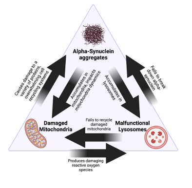
Research has brought us quite far in understanding some of the pathways that go awry in Parkinson’s and how specific cellular components might be malfunctioning. However, because all of these things are interconnected, we have a limited understanding of whether each cellular malfunction is a cause of neuron death, or a consequence of something else that negatively impacts the neuron’s health. For example, mitochondrial damage and the presence of protein aggregates can both lead to lysosomal damage24,26,27,28. Vice-versa, lysosomal damage can mean that malfunctioning mitochondria and alpha-synuclein are not being efficiently recycled by the cell19,29–31 (Figure 2). Such scenarios lead to a chicken-or-egg type controversy that makes it hard to define the origin of disease. Moreover, this article does not comprehensively list of all cellular processes associated with Parkinson’s; for example, the ability to move things from place to place within a neuron (called trafficking) is also thought to be dysfunctional in Parkinson’s32. Thus, while there are several cellular processes that are heavily involved in either causing or worsening the neuron death observed in Parkinson’s, the exact reason that neurons die is hard to pinpoint.
The good news is that every question we answer through research where the cause-and-effect of cellular malfunctions is clear brings us closer to understanding Parkinson’s pathology. In these scenarios, it is much easier to determine which problems can trigger specific cellular reactions than in actual diseased cells. In diseased cells, the precise timeline of cellular events is unknown, obscuring our cause-and-effect understanding of the different issues at hand. Fortunately, there are extensive research efforts currently in progress to better understand what’s happening to cause Parkinson’s. As complicated as the greater understanding of the disease pathology may be, many of the facts described here were not known as little as 10 years ago. With continued research, our understanding of the disease will only continue to improve, and once we understand what causes neuron death, we can work to create a way to prevent it.
References
1. O’Callaghan, C. & Lewis, S. J. G. Chapter Eighteen – Cognition in Parkinson’s Disease. in International Review of Neurobiology (eds. Chaudhuri, K. R. & Titova, N.) vol. 133 557–583 (Academic Press, 2017).
2. Péron, J., Dondaine, T., Le Jeune, F., Grandjean, D. & Vérin, M. Emotional processing in Parkinson’s disease: A systematic review. Mov. Disord. 27, 186–199 (2012).
3. Tysnes, O.-B. & Storstein, A. Epidemiology of Parkinson’s disease. J. Neural Transm. 124, 901–905 (2017).
4. Parkinson’s Disease Risk Factors and Causes. https://www.hopkinsmedicine.org/health/conditions-and-diseases/parkinsons-disease/parkinsons-disease-risk-factors-and-causes (2023).
5. Alpha-Synuclein – an overview | ScienceDirect Topics. https://www.sciencedirect.com/topics/neuroscience/alpha-synuclein.
6. Protein Aggregate – an overview | ScienceDirect Topics. https://www.sciencedirect.com/topics/neuroscience/protein-aggregate.
7. Stefanis, L. α-Synuclein in Parkinson’s Disease. Cold Spring Harb. Perspect. Med. 2, a009399 (2012).
8. Lamark, T. & Johansen, T. Aggrephagy: Selective Disposal of Protein Aggregates by Macroautophagy. Int. J. Cell Biol. 2012, 736905 (2012).
9. Hjerpe, R. et al. UBQLN2 Mediates Autophagy-Independent Protein Aggregate Clearance by the Proteasome. Cell 166, 935–949 (2016).
10. Longhena, F. et al. The Contribution of α-Synuclein Spreading to Parkinson’s Disease Synaptopathy. Neural Plast. 2017, 5012129 (2017).
11. Recasens, A. & Dehay, B. Alpha-synuclein spreading in Parkinson’s disease. Front. Neuroanat. 8, (2014).
12. Thorne, N. J. & Tumbarello, D. A. The relationship of alpha-synuclein to mitochondrial dynamics and quality control. Front. Mol. Neurosci. 15, (2022).
13. Brand, M. D., Orr, A. L., Perevoshchikova, I. V. & Quinlan, C. L. The role of mitochondrial function and cellular bioenergetics in ageing and disease. Br. J. Dermatol. 169, 1–8 (2013).
14. Marchi, S., Guilbaud, E., Tait, S. W. G., Yamazaki, T. & Galluzzi, L. Mitochondrial control of inflammation. Nat. Rev. Immunol. 23, 159–173 (2023).
15. Osellame, L. D., Blacker, T. S. & Duchen, M. R. Cellular and molecular mechanisms of mitochondrial function. Best Pract. Res. Clin. Endocrinol. Metab. 26, 711–723 (2012).
16. Tilokani, L., Nagashima, S., Paupe, V. & Prudent, J. Mitochondrial dynamics: overview of molecular mechanisms. Essays Biochem. 62, 341–360 (2018).
17. Dias, V., Junn, E. & Mouradian, M. M. The Role of Oxidative Stress in Parkinson’s Disease. J. Park. Dis. 3, 461–491 (2013).
18. Gao, X.-Y., Yang, T., Gu, Y. & Sun, X.-H. Mitochondrial Dysfunction in Parkinson’s Disease: From Mechanistic Insights to Therapy. Front. Aging Neurosci. 14, (2022).
19. Liu, J., Liu, W., Li, R. & Yang, H. Mitophagy in Parkinson’s Disease: From Pathogenesis to Treatment. Cells 8, 712 (2019).
20. Xiao, B., Kuruvilla, J. & Tan, E.-K. Mitophagy and reactive oxygen species interplay in Parkinson’s disease. Npj Park. Dis. 8, 1–13 (2022).
21. Bourdenx, M. & Dehay, B. What lysosomes actually tell us about Parkinson’s disease? Ageing Res. Rev. 32, 140–149 (2016).
22. Bourdenx, M., Bezard, E. & Dehay, B. Lysosomes and α-synuclein form a dangerous duet leading to neuronal cell death. Front. Neuroanat. 8, 83 (2014).
23. Shachar, T. et al. Lysosomal storage disorders and Parkinson’s disease: Gaucher disease and beyond. Mov. Disord. 26, 1593–1604 (2011).
24. Schneider, L. & Zhang, J. Lysosomal function in macromolecular homeostasis and bioenergetics in Parkinson’s disease. Mol. Neurodegener. 5, 14 (2010).
25. Spencer, B. et al. Beclin 1 Gene Transfer Activates Autophagy and Ameliorates the Neurodegenerative Pathology in α-Synuclein Models of Parkinson’s and Lewy Body Diseases. J. Neurosci. 29, 13578–13588 (2009).
26. Plotegher, N. & Duchen, M. R. Crosstalk between Lysosomes and Mitochondria in Parkinson’s Disease. Front. Cell Dev. Biol. 5, (2017).
27. Stepien, K. M. et al. Mechanisms of Mitochondrial Dysfunction in Lysosomal Storage Disorders: A Review. J. Clin. Med. 9, 2596 (2020).
28. Mazzulli, J. R., Zunke, F., Isacson, O., Studer, L. & Krainc, D. α-Synuclein–induced lysosomal dysfunction occurs through disruptions in protein trafficking in human midbrain synucleinopathy models. Proc. Natl. Acad. Sci. U. S. A. 113, 1931–1936 (2016).
29. Darios, F. & Stevanin, G. Impairment of Lysosome Function and Autophagy in Rare Neurodegenerative Diseases. J. Mol. Biol. 432, 2714–2734 (2020).
30. Doblado, L. et al. Mitophagy in Human Diseases. Int. J. Mol. Sci. 22, 3903 (2021).
31. Mani, S., Swargiary, G. & Chadha, R. Mitophagy impairment in neurodegenerative diseases: Pathogenesis and therapeutic interventions. Mitochondrion 57, 270–293 (2021).
32. Hunn, B. H. M., Cragg, S. J., Bolam, J. P., Spillantini, M.-G. & Wade-Martins, R. Impaired intracellular trafficking defines early Parkinson’s disease. Trends Neurosci. 38, 178–188 (2015).
Cover photo and figures were created by Julia Riley using BioRender.com.
Video was captured by Julia Riley.

I just want to compliment Julia Riley on her excellent PennNeuroknow column detailing current research in understanding Parkinson’s Disease. As a psychologist who needs to keep abreast of relevant neurological developments, I find that the PennNeuroknow series is excellent in communicating enough explanation, clearly-written, to advance my understanding without enmeshing me in poorly-explained technical details and jargon. This is the way science needs to be communicated to the public outside of the specialist confines of research groups. Science needs more of it. Thanks, and keep it coming.
LikeLiked by 1 person
Julia is delighted to receive this feedback, and our writers truly appreciate the compliment for their efforts to break down the brain for everyone to understand. Thank you!
LikeLike