April 29th, 2025
Written by: Andrew Nguyen
Imagine trying to navigate a city, with all of its intricate traffic patterns and interweaving streets, without using a map. This is surprisingly similar to how many neuroscientists have to study neural circuits. When Google Maps was first introduced, it allowed users to easily explore roads and highways to quickly find the best route from one location to another, all from their computer screens. Google Maps has also helped urban developers and engineers understand how traffic patterns work and what routes make it easiest to get from Point A to B, providing a lot of information about how people behave and the travel routes built to accommodate them. Now, imagine if there were a Google Maps of the brain! Instead of a roadmap of city streets, neuroscientists need a roadmap of neuronal connections (also known as synapses) that they can use to visualize and explore neural circuits that are important for performing certain behaviors, like feeding, sleeping, learning, and mating.
Building roadmaps of the brain
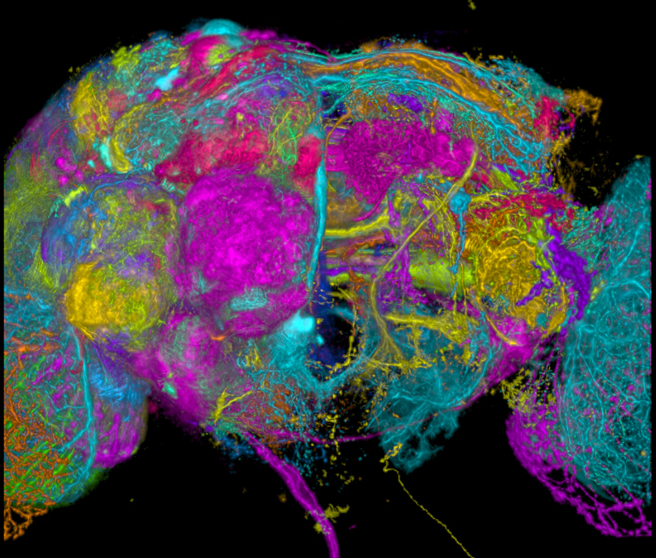
In October 2024, a collaborative group of dedicated neuroscientists known as the FlyWire Consortium published a landmark study sharing the world’s first publicly available, complete roadmap of the fruit fly brain called the “FlyWire connectome”1. The connectome is a reconstruction of how the neurons in the brain and nervous system are connected and interact. The FlyWire connectome builds the complete roadmap for the fly brain by reconstructing every neuron and their connections, taking the time to annotate (classify if they are excitatory/inhibitory, GABAergic, glutamatergic, etc.) every one. The tiny Drosophila melanogaster, or fruit fly, is an important animal model for neuroscientists studying how complex behaviors work (read all about fruit fly research here!). While the goal is to one day build roadmaps for the human brain, fruit flies are a great starting point because they still engage in many of the same complex behaviors that humans do, but have fewer neurons to map. As small as a sesame seed, the fruit fly brain contains around 140,000 neurons and around 54.5 million synapses (compared to humans’ ~86 billion neurons and over 100 trillion synapses). The fruit fly’s small but mighty group of neurons is responsible for coordinating complex behaviors like feeding, learning and memory, mating, social interactions, decision-making, navigation, and aggression. The fruit fly isn’t the first brain to get a roadmap, but it’s now the largest and most complex one we’ve uncovered, beating out the roundworm2 and the larval fruit fly3 for first place.
The creation of the fruit fly connectome is a huge scientific accomplishment. This collaborative group worked for close to a decade, with the most advanced computational and imaging technologies, to successfully create a complete roadmap of the fruit fly brain. Even though the fly brain is still far off from the billions of neurons in the human brain, the FlyWire connectome is a major step towards creating the connectome of the human brain. Camilo Guevara, a Neuroscience PhD Candidate at the University of Pennsylvania, explains why he’s excited about this development: “This is the first time that all the neurons and synapses of an animal with complex behaviors have been mapped. With this information, fly neuroscientists will be able to define circuits and generate hypotheses about the role of those circuits in behavior.”
The recipe for success
The FlyWire team was successful because of their patience, collaboration, and their advanced technology that allowed them to capture and annotate millions of images of the fly brains. The process required this team of researchers to use a technique called electron microscopy to take high-resolution images of fruit fly brain, little by little, at the nanometer scale (meanwhile a human hair is ~100,000 nanometers for scale!), and then develop new computational approaches with artificial intelligence and machine vision to align these images and reconstruct single 3D neurons. Based on data from other studies, the FlyWire team took it a step further to identify the connections between single neurons and annotate what type of synapse they are (excitatory/inhibitory), helping researchers understand their neural circuits even better4. They even classified each neuron’s shape and connectivity to better understand the diversity of neurons in the whole fly brain.
This incredible effort is already helping other neuroscientists in their pursuit to understand how the brain works. When the FlyWire connectome was released, the team worked with many other groups to demonstrate the value of their map. One team looked at how different types of neurons are connected to their neighbors. They found that some kinds of neurons receive a lot of inputs from other neurons and are thought to collect information, whereas others make more connections to other neurons and are thought to be more important for broadcasting that information across the brain5. Another team sought to define every neuron and synapse of the complex fly visual system, helping us understand how incredibly complicated visual inputs are relayed and represented in the brain6. These studies are just the beginning of how the FlyWire connectome could be used to further our understanding of the brain.
Shaping the future of neuroscience research
Now that the fly brain map has been available for six months, researchers like Guevara are beginning to use the connectome in new areas of research and offer powerful insight into how the brain works. He combines FlyWire with experiments in the lab to understand the connections between neurons that are important for sleep and circadian rhythms in the fruit fly brain and in the ventral nerve cord (which is essentially the fly’s version of a spinal cord). One of the many benefits of using the fruit fly is the vast number of genetic-based tools to visualize and activate/inhibit specific neurons in the brain and look at how these flies sleep as a result. Part of what makes FlyWire such a powerful tool is that it’s easy to use. “The platform for exploring the connection is open-source, user-friendly, and very intuitive,” said Guevara. “I learned [the essentials/basics] through hands-on practice, but they also offer great tutorials on their YouTube channel. You can even reach out with specific questions — the team is incredibly nice and responsive!”. The FlyWire connectome created a public, user-friendly resource for researchers to apply to their own work and has the potential to revolutionize the field of neuroscience!
As the field of neuroscience connectomics develops and the technologies to create more powerful tools evolves, future connectome resources should build off of previous work to learn from them and make them even better. While neuroscientists are already getting a lot of use out of FlyWire, there are still room for improvement. “One thing I would love to see is better integration with other [fly brain] connectome resources,” said Guevara, explaining that this could help create better annotations. “Making that connection easier would be incredibly helpful for experimental design.”
The FlyWire connectome represents a phenomenal breakthrough in neuroscience towards a complete reconstruction of the human brain. One major challenge is that the human brain is so much more complex with many more neurons and synapses to map. Imagine the computing power it must take to map out a smaller city compared to New York City – it’s the same for the fruit fly and the human brain. Many of the tools developed for the FlyWire connectome will be important building blocks to build the human connectome, but will require even more complex computational tools and imaging techniques. If putting the FlyWire connectome took close to a decade with the current technologies, mapping the human brain would be projected to finish in the next century. Luckily, researchers and engineers are collaborating to create the mouse connectome, which should be soon on their way with the fast-paced technological advancements, getting us even closer to creating Google Maps for the human brain.
References
- Dorkenwald, S., Matsliah, A., Sterling, A.R. et al. Neuronal wiring diagram of an adult brain. Nature 634, 124–138 (2024). https://doi.org/10.1038/s41586-024-07558-y
- Cook, S.J., Jarrell, T.A., Brittin, C.A. et al. Whole-animal connectomes of both Caenorhabditis elegans sexes. Nature 571, 63–71 (2019). https://doi.org/10.1038/s41586-019-1352-7
- Michael Winding et al. ,The connectome of an insect brain. Science 379, eadd9330 (2023). DOI: 10.1126/science.add9330
- Schlegel, P., Yin, Y., Bates, A.S. et al. Whole-brain annotation and multi-connectome cell typing of Drosophila. Nature 634, 139–152 (2024). https://doi.org/10.1038/s41586-024-07686-5
- Lin, A., Yang, R., Dorkenwald, S. et al. Network statistics of the whole-brain connectome of Drosophila. Nature 634, 153–165 (2024). https://doi.org/10.1038/s41586-024-07968-y
- Matsliah, A., Yu, Sc., Kruk, K. et al. Neuronal parts list and wiring diagram for a visual system. Nature 634, 166–180 (2024). https://doi.org/10.1038/s41586-024-07981-1
Cover image from Basisunus on Wikimedia Commons

Leave a comment