
November 3rd, 2026
Written by Emma Fischer
Head surgery, which includes both skull and brain surgery, dates back thousands of years. This long history has seen the techniques develop from drilling holes in the skull with stone tools to the use of robotics and nanotechnology in the modern neurosurgical room.1 The techniques and knowledge that underlie neurosurgery have undergone much development, but we would not be where we are today without the early scientists and doctors who pioneered brain surgery. Let’s unpack how ancient societies used early surgical techniques to provide care and how these techniques laid the groundwork for modern neurosurgery.
Ancient application of neurosurgery
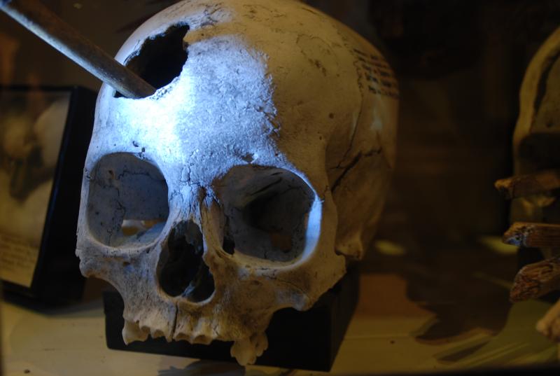
Although ancient civilizations had limited knowledge of the brain, doctors in these societies still tried to cure ailments like seizures, headaches, and mental illness.1,2 The earliest evidence we have of some of the treatments doctors attempted comes from skulls found across various continents dating back to 10,000 BCE!1,2 Some of these skulls had small holes in them that were created by ancient physicians in a practice known as trepanation. Tools such as obsidian or stone knives were used to drill these holes to treat a number of conditions, including headaches and traumatic head injuries.1,2 In Peru, trepanation was done using blades made from precious metals such as gold, though this metal is probably far too precious today to be used in the same way!1 These physicians reasoned that internal bleeding following head injuries caused a build up of intracranial pressure, or pressure pushing on the inside of the skull, and performed trepanation to relieve this pressure.2 While this may not have cured all ailments, and while it was certainly uncomfortable, creating a hole to allow bleeding from the brain to escape was very helpful in treating bleeding, and this concept is still used today. Archaeologists have found skulls with holes created from trepanation that show signs of injury healing after surgery, providing evidence that the treatment was successful in some of these cases!1
In order to advance neurosurgery beyond blindly drilling holes into the skull, science first needed to gain an understanding of how the head was connected to observed symptoms. Work from Greek and Roman scholars in the third century BCE used detailed studies of neuroanatomy to help the field realize that the brain served as the core of the nervous system.1,3
Over the next thousand years, physicians built on this body of work to develop further applications of trepanation and refine surgical tools, but the next crucial development came in the 19th century. Scientists had been conducting complex studies of neuroanatomy in order to better define the brain, and were able to describe distinct functions of different brain regions, or functional localization.1,3 For example, physician Pierre Paul Broca was able to link language deficits to a damaged region of the brain now known to be involved in speech production (see here for more fun case studies linking function to region).1 This allowed for more specific targeting of brain regions to address specific symptoms, and improved surgical accuracy and success.4 The advancement in the understanding of the brain’s central function in the body, combined with the increased use of anesthesia to reduce pain during surgery and better sanitation to limit infection, paved the way for centuries of unprecedented growth to drive complex and rapid advancements in neurosurgery.1,3, 4
Modern surgical developments
Medicine has seen an explosion in advancement since these foundational studies, and we now live in a time where innovations happen almost unbelievably quickly. Over the past sixty years, we have seen surgeons start to integrate fields such as robotics and physics to speed up the evolution of neurosurgery. The development of specialized microscopes allow surgeons to magnify even the smallest of blood vessels.5 These microscopes, referred to as operating microscopes, are used in many modern neurosurgeries to help surgeons see the complex (and tiny) system of nerves and tissue that make up our brains.5 This microscope allows neurosurgeons to repair nerves or veins and to remove tumors from the brain.5 Advanced operating microscopes have evolved to include robotic arms that can be controlled to more quickly and carefully localize the surgical target site, which makes surgery safer and more precise.
The understanding of functional localization has become much easier with time as we’re able to use tools for imaging the brain. Thanks to developments in the field of physics, we now have methods that allow us to view the brain in much more detail. For example, magnetic resonance imaging, or MRI, allows us to use powerful magnetic waves to better see the structure of the brain and blood flow between brain regions.6,7 As you can imagine, this method to see “into” the brain while someone is alive is a complete game changer for diagnosing, surgical targeting, and monitoring of conditions like stroke or brain cancer. Doctors can even use intraoperative MRI, or MRI done during surgery, to help them locate and remove brain tumors.8,9
Today, we have a vast and ever-expanding understanding of the brain and how to treat its disorders. The field of neurosurgery has evolved from early use of stone tools for trepanation to operating on a microscopic scale with the aid of imaging and microsurgical instruments. Though our understanding of neuroscience and neurosurgery is much changed since the first head surgeries were carried out, we owe our modern accomplishments to the work of scientists and physicians who came before us. Advancements in science and medicine are a collaborative effort, and we can look forward to much innovation in the future built on the hard work of scientists past and present.
References:
- Apuzzo, M. L., Liu, C. Y., Sullivan, D., & Faccio, R. A. (2007). Surgery of the human cerebrum—a collective modernity. Neurosurgery, 61(suppl_1), SHC-5.
- Bates, A. (2026). Beneath the surface: A historical exploration of trepanation practices across cultures and eras. Journal of Perioperative Practice, 36(3), 114-120.
- Nikova, A., & Birbilis, T. (2017). The basic steps of evolution of brain surgery. Maedica, 12(4), 297.
- Schijns, O. E., Hoogland, G., Kubben, P. L., & Koehler, P. J. (2015). The start and development of epilepsy surgery in Europe: a historical review. Neurosurgical review, 38(3), 447-461.
- Ma, L., & Fei, B. (2021). Comprehensive review of surgical microscopes: technology development and medical applications. Journal of biomedical optics, 26(1), 010901.
- Colen, R. R., Kekhia, H., & Jolesz, F. A. (2010). Multimodality intraoperative MRI for brain tumor surgery. Expert review of neurotherapeutics, 10(10), 1545-1558.
- Matthews, P. M., & Jezzard, P. (2004). Functional magnetic resonance imaging. Journal of Neurology, Neurosurgery & Psychiatry, 75(1), 6-12.
- Albayrak, B., Samdani, A. F., & Black, P. M. (2004). Intra-operative magnetic resonance imaging in neurosurgery. Acta neurochirurgica, 146(6), 543-557.
- Begley, S. L., McBriar, J. D., Pelcher, I., & Schulder M.. (2024). Intraoperative MRI: a review of applications across neurosurgical specialties. Neurosurgery 95, no. 3: 527-536.
No AI was used in this article.
Cover photo from Carlos Felipe Pardo on Flickr.

